
Courtesy: Dr Kshitij Chaudhury, Dr Kshitij Chaudhary is a fellowship-trained spine surgeon working at Sir HN Reliance Foundation Hospital Mumbai, India. He has been trained in USA at Harvard University’s Beth Israel Medical Center, Boston and Twin Cities Spine Center, Minneapolis. Web: http://spinemumbai.in/
Oncology Case Studies and Quiz- 2
Courtesy: Prof James Wittig Orthopaedic Oncologist Sarcoma Surgeon www.tumorsurgery.org James Wittig books:- Unknown Musculoskeletal Tumor Cases – Structured Clinical Review Overview This review summarizes important musculoskeletal tumor cases with emphasis on: Clinical presentation Imaging findings Histopathology Differential diagnosis Management principles A systematic approach using: Age Tumor location Matrix pattern MRI appearance Histology Immunohistochemistry is […]
Oncology Case Study and Quiz- 1
Courtesy: James Wittig Orthopaedic Oncologist Sarcoma Surgeon www.tumorsurgery.org James Wittig books:- Unknown Musculoskeletal Tumor Cases – Structured Review Overview These cases emphasize important principles in musculoskeletal oncology: Clinical presentation Imaging interpretation Histopathology correlation Differential diagnosis Management strategies Common themes: Age of patient Tumor location Matrix mineralization Biological behavior Histological pattern Case 11: Dedifferentiated Chondrosarcoma […]
Anatomy of Coracoid, Conoid and Coronoid
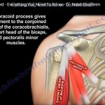
Courtesy: Prof Nabil Ebraheim, University of Toledo, Ohio, USA This video speaks about the coracoid, conoid, coronoid processes. Coracoid is the hook shaped bony process of scapula. It is considered as the surgeons lighthouse. It is safe to do surgical approaches lateral to coracoid process. The neuro vascular bundle lies medial to coracoid process. Three […]
Peri prosthetic Fractures: Advances in Management
COurtesy: Dr DD Tanna, Ashok Shyam, IORG, OrthoTV
Anatomy of Brachialis, Coracobrachialis and Brachioradialis

Courtesy: Prof Nabil Ebraheim, University of Toledo, Ohio, USA Overview The coracobrachialis, brachialis, and brachioradialis are commonly confused due to: Similar-sounding names Their role in flexion movements Their location along the anterior aspect of the upper limb Simple Memory Tip Think in proximal – distal order: Coracobrachialis – Shoulder (proximal) Brachialis – Arm (mid-region) Brachioradialis […]
Soft Tissue Sarcomas
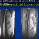
Courtesy: Prof James Wittig Orthopaedic Oncologist Sarcoma Surgeon www.tumorsurgery.org James Wittig books Soft Tissue Sarcomas Overview Soft tissue sarcomas are malignant tumors arising from mesenchymal tissues such as: Muscle Fat Fibrous tissue Blood vessels Peripheral nerves They may occur in: Extremities Pelvis Retroperitoneum Trunk Head and neck Common histological types include: Liposarcoma Undifferentiated pleomorphic […]
ORIF Proximal phalanx fracture of Ring finger
Courtesy: Dr Gregory Yanish MD, FACS Capital Orthopaedic and Sports Medicine, Clive, IOWA, USA
Benign Soft Tissue Tumours
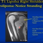
Courtesy: James Wittig Orthopaedic Oncologist Benign Soft Tissue Tumors: Structured Clinical Summary Overview Benign soft tissue tumors arise from soft tissues such as skin, subcutaneous tissue, muscle, and connective tissue surrounding bone. They do not originate from bone and do not metastasize. Common entities include lipoma, hemangioma, fibromatosis, myxoma, schwannoma, neurofibroma, giant cell tumor of […]
Open Reduction for DDH
Intramedullary Tibial Nailing- Surgical Technique
Small Round Blue Cell Tumours

Courtesy: Prof James Wittig Orthopaedic Oncologist Sarcoma Surgeon www.tumorsurgery.org James Wittig books Small Round Blue Cell Tumors of Bone Overview Group of tumors composed of: Small, round, blue-staining cells No matrix production (key feature) Resemble hematopoietic cells Include both benign and malignant entities Common Entities Benign Eosinophilic granuloma (Langerhans cell histiocytosis) Malignant Ewing sarcoma […]
Radiolucent Lesions of Bone
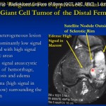
Courtesy: Prof James Wittig Orthopaedic Oncologist Sarcoma Surgeon www.tumorsurgery.org James Wittig books Radiolucent Bone Lesions: Structured Clinical Summary Overview Radiolucent bone lesions appear as lytic areas without internal mineralization on imaging. Common entities include giant cell tumor, aneurysmal bone cyst, unicameral bone cyst, eosinophilic granuloma, and non ossifying fibroma. Clinical interpretation depends on patient […]
Malignant Cartilage Tumours
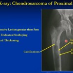
Courtesy: Dr James Wittig, Orthopaedic Oncologist Chondrosarcoma: Structured Clinical Summary Overview and Classification Chondrosarcoma is a malignant cartilage forming tumor with multiple histologic subtypes. It may arise primarily within normal bone or secondarily from preexisting lesions such as enchondroma or osteochondroma. Primary tumors constitute most cases, while secondary tumors are less common. Major subtypes include […]
Radiographic Densities
Courtesy: Learning Radiology