Courtesy: Prof James Wittig
Orthopaedic Oncologist
Sarcoma Surgeon
www.tumorsurgery.org
James Wittig books
Small Round Blue Cell Tumors of Bone
Overview
- Group of tumors composed of:
- Small, round, blue-staining cells
- No matrix production (key feature)
- Resemble hematopoietic cells
- Include both benign and malignant entities
Common Entities
- Benign
- Eosinophilic granuloma (Langerhans cell histiocytosis)
- Malignant
- Ewing sarcoma
- Lymphoma
- Multiple myeloma / Plasmacytoma
- Metastatic neuroblastoma
- Metastatic small cell carcinoma
- Rhabdomyosarcoma (rare in bone)
1. Eosinophilic Granuloma (Langerhans Cell Histiocytosis)
Nature
- Benign proliferation of Langerhans cells
- Often associated with:
- Eosinophils
- Lymphocytes
- Plasma cells
- Considered immune dysregulation, not true neoplasm
Epidemiology
- Age: 5–15 years (most common)
- Male predominance (2:1)
Types
- Solitary lesion (70%)
- Multifocal disease
- Disseminated forms:
- Hand-Schüller-Christian disease
- Triad:
- Lytic bone lesions
- Exophthalmos
- Diabetes insipidus
- Triad:
- Letterer-Siwe disease
- <1 year
- Highly fatal
- Hand-Schüller-Christian disease
Common Sites
- Flat bones (70%)
- Skull
- Pelvis
- Long bones (less common)
Clinical Features
- Pain + swelling
- Mild fever
- Peripheral eosinophilia (5–10%)
Radiology
- Variable:
- Geographic (benign)
- Moth-eaten / permeative (aggressive)
- Features:
- Onion-skin periosteal reaction
- Possible sequestrum (“button sequestrum”)
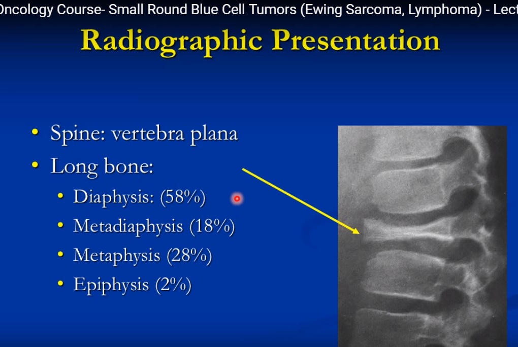
- Spine: Vertebra plana (classic)
MRI
- Marrow replacement
- T2 hyperintensity
- May show soft tissue mass
Pathology
- Langerhans cells – coffee-bean nuclei
- Birbeck granules (tennis racket appearance)
- Immunostains:
- CD1a+
- S100+
- Vimentin+
Treatment
- Observation (many resolve)
- Curettage + bone graft (if fracture risk)
- Intralesional steroids
- Rare: radiation
2. Ewing Sarcoma
Key Concept
- Most important malignant small round blue cell tumor
- Always considered micrometastatic at presentation
Epidemiology
- Age: 10–25 years
- Slight male predominance
- Rare in African ancestry
Genetics
- t(11;22) – EWS-FLI1 fusion protein
Common Sites
- Femur
- Humerus
- Pelvis
- Ribs
Diaphyseal origin (key exam point)
Clinical Features
- Pain + swelling
- Fever, anemia, elevated ESR (may mimic infection)
- Pathological fracture (~10%)
Radiology
- Permeative / moth-eaten lesion
- Onion-skin periosteal reaction
- Large soft tissue mass (90%)
- No matrix production
MRI
- Extensive marrow involvement
- Large soft tissue component
- Extends beyond what X-ray shows
Bone Scan
- Intense uptake
Pathology
- Uniform monotonous small round blue cells
- Minimal cytoplasm
- Glycogen-rich – PAS positive
- Immunostains:
- CD99+
- Vimentin+
- HBA71+
Treatment
- Multi-agent chemotherapy (essential)
- Limb-sparing surgery (preferred)
- Radiation (selected cases)
Prognosis
- Localized disease:
- ~65% 5-year survival
- Metastatic disease:
- 15–30% survival
- Worse:
- Pelvic tumors
3. Primary Lymphoma of Bone
Definition
- Lymphoma arising in bone without systemic disease for 6 months
Epidemiology
- Usually >40 years
- Rare in children
Common Sites
- Femur
- Pelvis
- Humerus
Clinical Features
- Dull pain
- Palpable mass
- Pathological fracture (25%)
Radiology
- Permeative / moth-eaten
- Sometimes normal X-ray
- Soft tissue mass common
MRI
- Marrow replacement
- Soft tissue extension without cortical destruction
Pathology
- Mixed cell population:
- Small lymphocytes
- Large malignant B cells
- Variable size cells (unlike Ewing)
Markers
- CD20+
- CD45+
- Reticulin+
- Leukocyte antigen+
Treatment
- Chemotherapy ± radiotherapy
- Surgery only for stabilization
4. Multiple Myeloma / Plasmacytoma
Nature
- Malignant proliferation of plasma cells
Types
- Multiple myeloma (systemic)
- Solitary plasmacytoma
Epidemiology
- Age: >50 years
- Most common primary bone malignancy
Common Sites
- Spine
- Pelvis
- Skull
- Ribs
- Proximal femur
Clinical Features
- Bone pain
- Anemia
- Hypercalcemia
- Renal failure
- Pathological fractures
Investigations
- SPEP: M protein spike
- Urine: Bence Jones protein
- ESR
Radiology
- Punched-out lytic lesions
- No sclerosis
- Diffuse osteopenia
Bone Scan
- May be negative
Pathology
- Plasma cells:
- Clock-face nucleus
- Perinuclear halo
Special Syndrome
- POEMS syndrome
- Polyneuropathy
- Organomegaly
- Endocrinopathy
- Monoclonal gammopathy
- Skin changes
Treatment
- Chemotherapy
- Stem cell transplant
- Radiotherapy (localized disease)
- Surgery (fracture fixation)
High-Yield Comparison Table
| Feature | Eosinophilic Granuloma | Ewing Sarcoma | Lymphoma | Myeloma |
|---|---|---|---|---|
| Age | 5–15 | 10–25 | >40 | >50 |
| Nature | Benign | Malignant | Malignant | Malignant |
| Matrix | None | None | None | None |
| Cells | Langerhans | Uniform SRBCT | Mixed cells | Plasma cells |
| Key Feature | Vertebra plana | Onion-skin + soft tissue mass | Mixed infiltrate | Punched-out lesions |
| Marker | CD1a, S100 | CD99 | CD20 | Ig spike |
Leave a Reply