Courtesy: Dr James Wittig,
Orthopaedic Oncologist
Chondrosarcoma: Structured Clinical Summary
Overview and Classification
- Chondrosarcoma is a malignant cartilage forming tumor with multiple histologic subtypes.
- It may arise primarily within normal bone or secondarily from preexisting lesions such as enchondroma or osteochondroma.
- Primary tumors constitute most cases, while secondary tumors are less common.
- Major subtypes include conventional intramedullary, clear cell, mesenchymal, dedifferentiated, and periosteal chondrosarcoma.
Conventional Chondrosarcoma
- Most common subtype, usually affecting adults older than forty years.
- Common locations include pelvis, proximal femur, distal femur, proximal humerus, ribs, and scapula.
- Patients typically present with pain, sometimes associated with swelling; pathological fracture is uncommon.
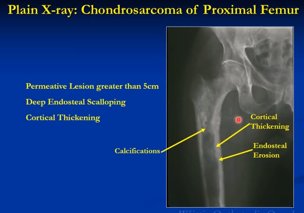
- Radiographs show metaphyseal or diaphyseal lesions with ring and arc calcifications and endosteal scalloping.
- Malignant features include cortical thickening, deep endosteal scalloping, cortical destruction, bone expansion, and soft tissue extension.
Distinguishing Enchondroma from Low Grade Chondrosarcoma
- Low grade chondrosarcoma typically occurs in older patients and presents with pain related to the lesion.
- Lesions are usually larger than five centimeters with deeper endosteal scalloping.
- Bone scan uptake is usually greater than the anterior superior iliac spine.
- Soft tissue extension, cortical thickening, and periosteal reaction favor malignancy.
Histologic Grading
- Grade one tumors resemble benign cartilage with low cellularity and minimal atypia but demonstrate entrapment of host trabeculae.
- Grade two tumors show increased cellularity, nuclear enlargement, binucleation, and occasional mitoses.
- Grade three tumors demonstrate marked pleomorphism, high cellularity, spindle morphology, and frequent mitoses.
- Higher grade correlates with increased risk of metastasis and aggressive behavior.
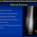
Dedifferentiated Chondrosarcoma
- Characterized by abrupt transition between low grade cartilage tumor and high grade noncartilaginous sarcoma.
- Common high grade components include osteosarcoma, fibrosarcoma, and undifferentiated pleomorphic sarcoma.
- Highly aggressive tumor with poor prognosis and high metastatic rate.
- Radiographs show biphasic pattern with calcified cartilage component and adjacent destructive lytic area with soft tissue mass.
Secondary Chondrosarcoma
- Arises from osteochondroma or enchondroma, often in pelvis, scapula, ribs, or proximal femur.
- Malignant transformation suspected with cartilage cap thickness greater than two centimeters on imaging.
- Other concerning features include growth after skeletal maturity, cortical destruction, and increasing pain.
Clear Cell Chondrosarcoma
- Rare low to intermediate grade tumor usually arising in the epiphysis of long bones.
- Most common sites include proximal femur and proximal humerus.
- Radiographs show lytic epiphyseal lesion with mild sclerosis and minimal calcification.
- Histology shows clear cytoplasm rich in glycogen and S100 positivity.
Mesenchymal Chondrosarcoma
- High grade tumor composed of small round cells with islands of malignant cartilage.
- Occurs in young adults and may arise in bone or soft tissue.
- Common sites include femur, ribs, pelvis, jaw, and spine.
- Characterized by aggressive behavior with high metastatic potential.
Periosteal Chondrosarcoma
- Surface tumor arising from periosteum and eroding cortex without medullary involvement.
- Common in femur and humerus and usually presents as painless swelling.
- Radiographs show saucerization of cortex with soft tissue mass and chondroid calcification.
Treatment and Prognosis
- Surgical resection is the primary treatment for all chondrosarcoma subtypes.
- Limb sparing surgery is preferred when feasible, though amputation may be required in advanced cases.
- Chemotherapy and radiotherapy are generally ineffective except in selected high grade or unresectable tumors.
- Prognosis depends mainly on histologic grade, with higher grade tumors showing worse outcomes.
Leave a Reply