Courtesy: Medical Lectures Made Easy
Overview of Bone and Cartilage Tumors: Benign and Malignant Lesions
Introduction
- Bone and cartilage tumors include a wide spectrum of benign and malignant neoplasms affecting hard tissues.
- These tumors vary in age distribution, anatomical location, imaging features, histology, and treatment.
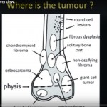
Benign Bone and Cartilage Tumors
Osteoma
- Benign bone tumor most commonly involving skull and facial bones.
- May obstruct paranasal sinuses and lead to infection or breathing difficulty.
- Associated with Gardner syndrome.
Osteoid Osteoma
- Benign bone forming tumor commonly affecting the cortex of long bones.
- Typically occurs in young patients, more often in males.
- Commonly arises in the diaphysis.
- Radiographs show a small central nidus surrounded by reactive sclerosis.
- Usually less than two centimeters in size.
- Clinically presents with nocturnal pain relieved by anti inflammatory medication.
- Histology shows immature bone with osteoblasts and osteoclasts without atypia.
- Treatment includes observation or radiofrequency ablation when symptomatic.
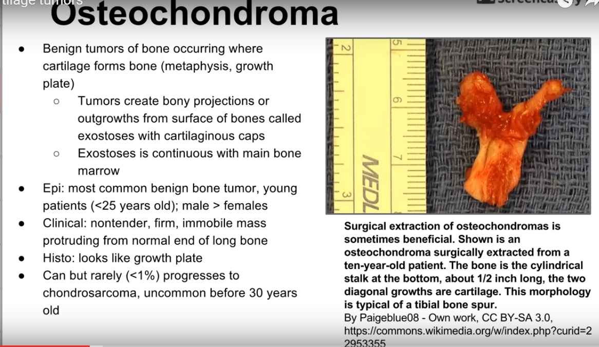
Osteochondroma
- Most common benign bone tumor arising from the metaphysis near the growth plate.
- Forms a bony outgrowth with a cartilage cap.
- The marrow cavity is continuous with the parent bone.
- Usually occurs in young individuals.
- Typically presents as a painless palpable mass.
- Rare malignant transformation to chondrosarcoma may occur.
- Treatment is observation unless symptomatic.
Nonossifying Fibroma
- Fibrous cortical lesion seen in children and adolescents.
- Commonly involves the metaphysis of long bones such as the femur and tibia.
- Radiographs show eccentric, lobulated lesions with a thin sclerotic rim.
- Usually asymptomatic and discovered incidentally.
- Histology shows fibrous tissue with foamy histiocytes.
- Typically resolves spontaneously without treatment.
Giant Cell Tumor
- Usually arises in the epiphysis after skeletal maturity.
- Commonly occurs around the knee.
- Often benign but locally aggressive.
- Radiographs show a soap bubble appearance without sclerosis.
- Histology shows multinucleated giant cells with uniform stromal cells.
- Treatment includes curettage with grafting or cementation.
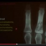
Chondroma
- Benign cartilage tumor arising within bone.
- Most often affects the small bones of the hands and feet.
- Radiographs show central calcifications with rings and arcs pattern.
- Usually asymptomatic and found incidentally.
- Rare malignant transformation to chondrosarcoma.
Malignant Bone and Cartilage Tumors
Osteosarcoma
- Highly aggressive malignant bone tumor with osteoblastic differentiation.
- Typically arises in the metaphysis of long bones around the knee.
- Common in adolescents with a second peak in older adults.
- Risk factors include prior radiation, Paget disease, and genetic syndromes.
- Radiographs show sunburst pattern and Codman triangle.
- Histology demonstrates malignant cells producing osteoid.
- Treatment includes chemotherapy and surgical resection.
Ewing Sarcoma
- Malignant tumor composed of small round blue cells.
- Commonly involves diaphysis or metadiaphysis of long bones.
- Associated with chromosome translocation involving eleven and twenty two.
- Occurs mainly in children and adolescents.
- Radiographs show onion skin periosteal reaction.
- Responds well to chemotherapy combined with local treatment.
Chondrosarcoma
- Malignant cartilage forming tumor typically affecting older adults.
- Often arises in the pelvis, spine, or scapula.
- Radiographs show stippled calcification and cortical involvement.
- Histology shows increased cellularity and atypia compared with benign cartilage tumors.
- Treatment primarily involves wide surgical resection since chemotherapy and radiation are less effective.
Summary
- Bone and cartilage tumors range from benign incidental lesions to aggressive malignancies.
- Accurate diagnosis depends on clinical, radiographic, and histological correlation.
Appropriate treatment varies from observation to multimodal oncologic therapy
Leave a Reply