-Classifications
Outerbridge Classification of Cartilage defects

? Courtesy: Christopher McCrum, UT SouthWestern, Dallas, Texas, USA Outerbridge Bentley Classification: Grade I lesion: articular surfaces that are swollen, soft, and in some cases blistered. Grade II lesions: characterized by articular fissures and clefts with diameters <1 cm. Grade III lesions: deep fissures extending to subchondral bone with diameters >1 cm. Grade IV lesions: […]
Stulberg Classification in Perthe’s Disease

Courtesy: Sally Hobson, Hull ROyal Infirmary, Hull, UK Stulberg Classification (Based on Femoral Head Shape and Congruence) This classification evaluates the shape of the femoral head and its congruence with the acetabulum on radiographs after healing. Type I – Spherically Congruent (Normal) Femoral head: Completely normal and spherical. Acetabulum: Normal. Joint congruence: Perfect congruence between […]
Herring Classification for Perthe’s Disease
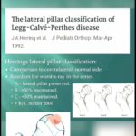
? Courtesy: Dr Sally Hobson, Hull Royal Infirmary, Hull, UK Herring (Lateral Pillar) Classification This classification is based on the preservation of the lateral pillar of the femoral head epiphysis on anteroposterior radiographs during the fragmentation stage. Type A Lateral pillar height: 100% preserved (no loss of height). Femoral head: Maintains normal height and structure. […]
Classification of Throwing Phases in Athletes

Courtesy: Mathew Binkley MD, Assistant Professor, University of Buffalo, USA The six phases of throwing: wind-up stride (early cocking) late cocking acceleration deceleration follow through
Wassel Classification of Thumb Polydactyly

Courtesy: Rishi Dhir, FRCS Orth, UK Wassel Classification of Thumb Polydactyly type I describes a bifid distal phalanx type II, duplicated distal phalanx type III, bifid proximal phalanx type IV, duplicated proximal phalanx type V, bifid metacarpal type VI, duplicated metacarpal type VII, triphalangism
Brumback Classification of Femoral Head Fractures

? Orthopaedic Principles Shorts Channel Type 1: posterior hip dislocation with fracture of femoral head involving inferomedial (non weight bearing) portion of femoral head. Type 1A, with minimal or no fracture of acetabular rim and stable hip joint after reduction. Type 1B, with significant acetabular fracture and hip joint instability. Type 2: posterior hip dislocation […]
Salter Harris Classification of Epiphyseal Injuries
Courtesy: Prof Nabil Ebraheim, University of Toledo, Ohio, USA GROWTH PLATE ANATOMY AND ITS FUNCTIONS INTRODUCTION The growth plate, also known as the physis, is the cartilaginous portion at the ends of long bones where longitudinal growth of the bone takes place. This region of bone is characterized by high metabolic activity and is under […]
Classification of Pelvic Fractures
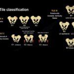
Courtesy: Prof Nabil Ebraheim, University of Toledo, Ohio, USA Pelvic Fracture Classification: Young–Burgess and Tile Systems Introduction Pelvic fracture classification is essential to determine: Severity of injury Pelvic stability Risk of hemorrhage Need for surgical intervention Clinical Importance Initial pelvic X-ray and patient condition help classify injuries as: Stable (simple) Unstable (life-threatening) ? […]
Patterson Classification of Constriction Band Syndrome
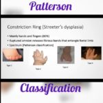
Class 1 simple constriction ring Class 2 associated deformity of the distal part +/- lymphedema. Class 3 involves distal fusions like syndactyly/acrodactyly Class 4 involved intrauterine amputation due to the constriction Classic Ref: Patterson T. Congenital ring constrictions. Br J Plast Surg. 1961;69:532–569
Supination-Adduction in Lauge Hansen Classification

Courtesy: Dr Glass DPM Supination–Adduction (SA) Injury (Lauge–Hansen Classification of Ankle Fractures) Overview Supination–Adduction (SA) injuries are a distinct pattern of ankle fractures described in the Lauge–Hansen classification, based on mechanism of injury. Key Concept Injury pattern follows a predictable sequence depending on the applied force Mechanism of Injury Position of Foot Supinated foot Applied […]
Danis Weber Classification of Ankle fractures

Courtesy: Prof Nabil Ebraheim, University of Toledo, Ohio, USA Fibular fracture that is more proximal indicates an increased risk of syndesmotic disruption and ankle instability. This classification is based on the level of fibula fracrture 1. TYPE A Internal rotation and adduction injury. Fracture of the fibula below the level of tibial plafond. Usually an […]
Cierny and Mader Classification For Osteomyelitis

Anatomical type: • Type I, Medullary: osteomyelitis is confined to the medullary cavity of the bone. Hematogenous osteomyelitis and infections in the presence of an intramedullary rod are examples of this stage. • Type II, Superficial: involves only the cortical bone and usually originates from a direct inoculation or a contiguous focus infection. Typically seen […]
University of Texas Classification for Diabetic Foot

University of Texas Classification Stage Description A No infection or Ischemia B Infection present C Ischemia present D Infection and Ishcemia present Grading Description 0 Epithelialised wound 1 Superficial wound 2 Wound penetrates to tendon/capsule 3 Wound penetrates to bone/joint
Radial Club Hand Classification
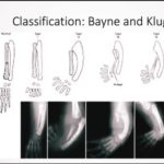
Bayne and Klug Classification Type I Short Distal Radius (Hypoplastic/Absent distal radial epiphysis) Type II Hypoplastic Radius (Deficient radial epiphyses, distal and proximal) Type III Partial Absence(Present Proximally) Type IV Complete Absence (Most Common) Full Video: https://youtu.be/2toAbtsNZTQ Ref: Long-term review of the surgical treatment of radial deficiencies Loui G.Bayne M.D. Mark S.Klug M.D. The Journal […]