Courtesy: Prof James Wittig
Orthopaedic Oncologist
Sarcoma Surgeon
www.tumorsurgery.org
James Wittig books:-
Unknown Musculoskeletal Tumor Cases – Structured Clinical Review
Overview
This review summarizes important musculoskeletal tumor cases with emphasis on:
- Clinical presentation
- Imaging findings
- Histopathology
- Differential diagnosis
- Management principles
A systematic approach using:
- Age
- Tumor location
- Matrix pattern
- MRI appearance
- Histology
- Immunohistochemistry
is essential for diagnosis.
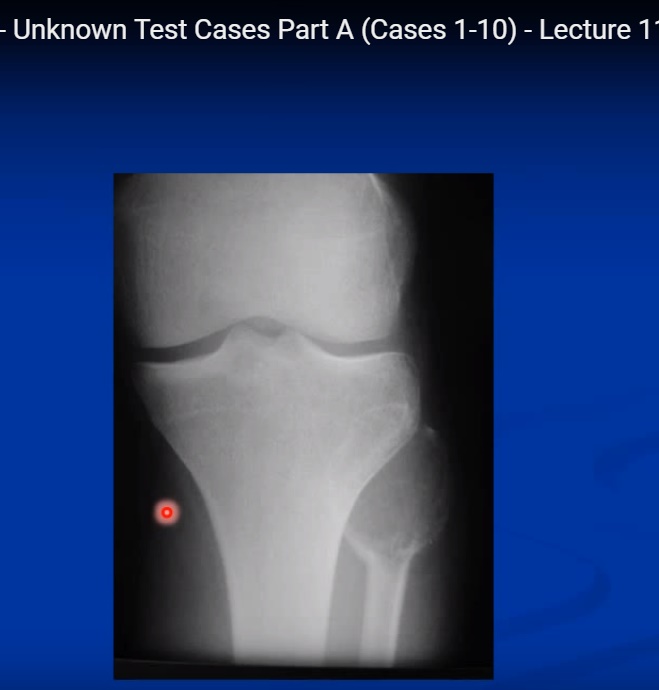
Case 1: Pigmented Villonodular Synovitis (PVNS)
Clinical Features
- Middle-aged woman
- Chronic anterior knee pain
- Swelling and recurrent effusion
MRI Findings
- Diffuse synovitis
- Low-signal foci from hemosiderin deposition
Histology
- Synovial fronds
- Giant cells
- Foamy histiocytes
- Fibrous tissue
- Hemosiderin-laden macrophages
Types
Diffuse Type
- More aggressive
- Higher recurrence
Nodular Type
- Lower recurrence after excision
Treatment
- Synovectomy
- Diffuse disease may require:
- Staged surgery
- Adjuvant radiotherapy
Case 2: Giant Cell Tumor (GCT) of Bone
Clinical Features
- Young skeletally mature adult
- Mild pain and swelling
- Common around knee
Imaging Findings
- Geographic expansile lytic lesion
- Cortical thinning
- Internal trabeculations
Histology
Characteristic finding:
- Uniform giant cells mixed with stromal cells having similar nuclei
Important Features
- Benign but locally aggressive
- Usually epiphyseal lesion after physeal closure
Treatment
- Intralesional curettage
- Local adjuvants
- Bone graft/cement reconstruction
- Wide resection in selected sites
Case 3: Adamantinoma
Clinical Features
- Adolescent male
- Long-standing tibial bowing
Imaging Findings
- Expansile tibial lesion
- Cortex + medullary involvement
Histology
- Biphasic tumor:
- Spindle cells
- Epithelial nests
Immunohistochemistry:
- Cytokeratin positive
Diagnosis
- Low-grade malignant tumor
Treatment
- Wide resection
- Reconstruction
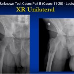
Case 4: Plasma Cell Neoplasm
Clinical Features
- Older adult
- Hip pain
- Proximal femoral lytic lesion
Histology
- Uniform plasma cells
- Clock-face chromatin
- Perinuclear hof/halo
Differential Diagnosis
- Multiple myeloma
- Solitary plasmacytoma
Workup
- Serum electrophoresis
- Urine electrophoresis
- Bone marrow examination
Management
Depends on systemic disease involvement.
Orthopedic indication:
- Fixation for impending/pathological fracture
Case 5: Conventional Intramedullary Osteosarcoma
Clinical Features
- Adolescent patient
- Progressive knee pain
- Elevated alkaline phosphatase
Imaging Findings
- Permeative lesion
- Cloud-like osteoid matrix
- Cortical destruction
Histology
- Pleomorphic malignant cells
- Lace-like osteoid production
Standard Treatment
- Neoadjuvant chemotherapy
- Wide surgical resection
- Adjuvant chemotherapy
Prognostic Factors
- Stage
- Metastases
- Response to chemotherapy
Case 6: Myxoid Liposarcoma
Clinical Features
- Adult patient
- Painless enlarging intramuscular mass
MRI Findings
- High T2 signal
- Myxoid appearance
Histology
- Lipoblasts
- Myxoid matrix
- Delicate capillary network
Treatment
- Wide resection
- Postoperative radiotherapy commonly used
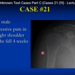
Case 7: Chondroblastoma
Clinical Features
- Adolescent
- Shoulder pain
Imaging Findings
- Well-defined epiphyseal lesion
- Internal calcification
- Surrounding edema
Histology
Classic findings:
- Chondroblasts
- Coffee-bean nuclei
- Chicken-wire calcification
Treatment
- Curettage
- Bone grafting
Complication
- Local recurrence
Case 8: Angiosarcoma
Clinical Features
- Rapidly enlarging painless arm mass
Histology
- Malignant endothelial proliferation
- Marked atypia
Immunohistochemistry
Positive vascular markers:
- CD31
- CD34
Treatment
- Surgery
- Chemotherapy
- Radiotherapy
Case 9: Ewing Sarcoma
Clinical Features
- Pelvic lesion
- Small round blue cell tumor
Histology
- Monotonous small round cells
- No matrix production
Immunohistochemistry
- CD99 positive
Cytogenetics
Characteristic translocation:
- t(11;22)
Treatment
- Multi-agent chemotherapy
- Surgical resection
- Radiotherapy in selected cases
Case 10: Undifferentiated Pleomorphic Sarcoma
Clinical Features
- Older adult
- Large intramuscular thigh mass
Imaging Findings
- Deep soft tissue lesion
- Necrosis and hemorrhage
Histology
- Pleomorphic spindle cells
- Storiform pattern
Treatment
- Wide resection
- Radiotherapy
- Selective chemotherapy
High-Yield Imaging Clues
| Imaging Feature | Suggestive Diagnosis |
|---|---|
| Hemosiderin low signal on MRI | PVNS |
| Epiphyseal lesion with chicken-wire calcification | Chondroblastoma |
| Cloud-like osteoid matrix | Osteosarcoma |
| Myxoid high T2 lesion | Myxoid liposarcoma |
| Expansile epiphyseal lytic lesion | Giant cell tumor |
| Small round blue cell tumor | Ewing sarcoma |
Important Histology Pearls
| Tumor | Characteristic Histology |
|---|---|
| Giant cell tumor | Uniform giant cells |
| Chondroblastoma | Chicken-wire calcification |
| Schwannoma | Antoni A/B + Verocay bodies |
| Osteosarcoma | Malignant osteoid |
| Fibrosarcoma | Herringbone pattern |
| Leiomyosarcoma | Cigar-shaped nuclei |
| Plasma cell neoplasm | Clock-face chromatin |
Important Management Principles
- Biopsy before definitive treatment
- Limb salvage preferred when feasible
- Wide margins critical for malignant tumors
- Radiotherapy important for local control in soft tissue sarcoma
- Chemotherapy essential in:
- Osteosarcoma
- Ewing sarcoma
- Rhabdomyosarcoma
Key Exam Pearls
- Giant cell tumor occurs after skeletal maturity
- Chondroblastoma is epiphyseal in adolescents
- PVNS contains hemosiderin-laden macrophages
- Ewing sarcoma is CD99 positive
- Osteosarcoma produces malignant osteoid
- Adamantinoma commonly affects tibial diaphysis
- Myxoid liposarcoma has high T2 MRI signal
- Plasma cell tumors require systemic workup
Leave a Reply