Courtesy: James Wittig,Orthopaedic oncologist
Unknown Musculoskeletal Tumor Cases: Diagnostic Approach and Key Lessons
Overview
- This session reviews unknown musculoskeletal tumor cases using a structured diagnostic approach.
- Each case integrates clinical presentation, radiographic interpretation, histology, and treatment principles.
- The approach is particularly useful for examination preparation and clinical decision making.
General Diagnostic Approach
- Begin with clinical details including age, symptoms, and duration.
- Analyze radiographs for lesion location, pattern of bone destruction, matrix mineralization, and periosteal reaction.
- Use magnetic resonance imaging to assess intraosseous extent and soft tissue involvement.
- Correlate imaging findings with histopathology.
- Formulate differential diagnosis before confirming the final diagnosis.
Case Based Highlights
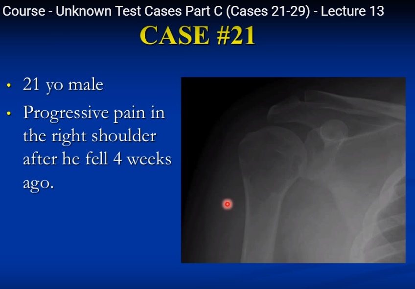
Case One: Low Grade Cartilage Tumor of Proximal Humerus
- Young adult with shoulder pain and metaphyseal lesion extending into epiphysis.
- Imaging showed lobulated lesion with cartilage signal characteristics and extraosseous extension.
- Histology revealed hypercellular hyaline cartilage with atypia.
- Diagnosis: Grade one conventional intramedullary chondrosarcoma.
- Management typically involves surgical resection; intralesional procedures may be considered in selected cases.
Case Two: Dedifferentiated Cartilage Tumor with Pathological Fracture
- Elderly patient with pathological fracture and mixed lytic and mineralized lesion.
- Radiographs suggested cartilage tumor with aggressive component.
- Histology demonstrated low grade cartilage tumor adjacent to high grade sarcoma.
- Diagnosis: Dedifferentiated chondrosarcoma.
- Management includes wide surgical resection; prognosis is generally poor.
Case Three: Eccentric Proximal Tibial Lesion
- Young adult with knee pain and eccentric metaphyseal lesion extending into epiphysis.
- Radiographs showed geographic destruction and soft tissue extension.
- Histology demonstrated multinucleated giant cells with matching mononuclear cells.
- Diagnosis: Giant cell tumor of bone.
- Treatment includes curettage with adjuvant therapy and reconstruction.
Case Four: Aggressive Distal Femoral Lesion in Adult
- Adult patient with permeative lesion and soft tissue mass.
- Histology showed pleomorphic spindle cell sarcoma with storiform pattern.
- Diagnosis: Undifferentiated pleomorphic sarcoma of bone.
- Treatment includes chemotherapy and wide surgical resection.
Case Five: Expansile Distal Tibial Lesion in Child
- Child with metaphyseal expansile lesion showing internal septations.
- MRI demonstrated fluid fluid levels.
- Histology showed blood filled cystic spaces and reactive bone formation.
- Diagnosis: Aneurysmal bone cyst.
- Treatment includes curettage and bone grafting.
Case Six: Epiphyseal Lesion in Child
- Skeletally immature patient with knee pain and epiphyseal lesion.
- MRI demonstrated surrounding edema and variable signal intensity.
- Histology showed chondroblasts with coffee bean nuclei and chicken wire calcification.
- Diagnosis: Chondroblastoma.
- Treatment includes curettage and bone grafting.
Case Seven: Small Round Cell Tumor in Child
- Young child with pain, swelling, and systemic symptoms.
- Radiographs showed aggressive lesion with sclerosis.
- Histology revealed monotonous small round blue cells.
- Diagnosis: Ewing sarcoma.
- Management includes chemotherapy with surgery and selective use of radiation.
Case Eight: Sclerotic Proximal Femoral Lesion in Older Adult
- Elderly patient with predominantly sclerotic lesion.
- Histology showed mixed lymphoid population with large atypical cells.
- Diagnosis: Lymphoma of bone.
- Treatment includes chemotherapy and radiation.
Case Nine: Aggressive Metaphyseal Lesion in Adolescent
- Teenager with enlarging knee mass.
- Radiographs showed mixed lytic and sclerotic lesion with soft tissue mass and periosteal reaction.
- Histology demonstrated malignant osteoid production.
- Diagnosis: High grade osteosarcoma.
- Management includes chemotherapy and surgical resection.
Key Learning Points
- Patient age and lesion location significantly narrow differential diagnosis.
- Benign tumors often show geographic margins and sclerotic rims.
- Malignant lesions typically demonstrate permeative destruction and soft tissue extension.
- Histological correlation is essential for definitive diagnosis.
- Treatment varies widely depending on tumor type and grade.
Summary
- A systematic approach improves diagnostic accuracy in musculoskeletal tumors.
- Integration of clinical, radiological, and pathological findings is essential for optimal management.
Leave a Reply