Courtesy: James Wittig
Orthopaedic Oncologist
Sarcoma Surgeon
www.tumorsurgery.org
James Wittig books:-
Unknown Musculoskeletal Tumor Cases – Structured Review
Overview
These cases emphasize important principles in musculoskeletal oncology:
- Clinical presentation
- Imaging interpretation
- Histopathology correlation
- Differential diagnosis
- Management strategies
Common themes:
- Age of patient
- Tumor location
- Matrix mineralization
- Biological behavior
- Histological pattern
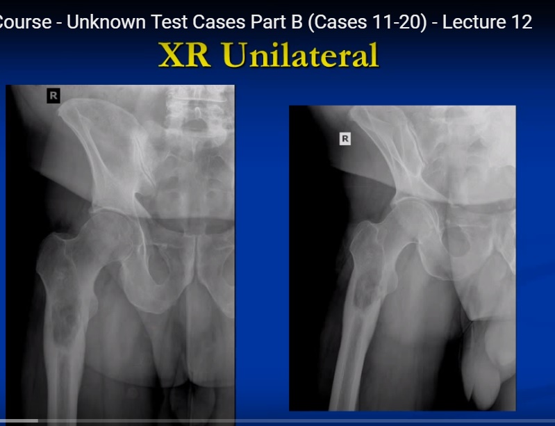
Case 11: Dedifferentiated Chondrosarcoma
Clinical Features
- Older adult
- Progressive hip pain
- Proximal femoral lesion
Imaging Findings
Radiographs showed:
- Lytic destructive lesion
- Cortical thickening
- Cartilage-type calcification
- Ring-and-arc pattern
Histology
Characteristic finding:
- Abrupt transition from low-grade cartilage tumor to high-grade sarcoma
Common dedifferentiated components:
- Undifferentiated pleomorphic sarcoma
- Osteosarcoma
- Fibrosarcoma
Prognosis
- Highly aggressive
- Poor prognosis
Treatment
- Wide resection
- Reconstruction
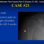
Case 12: Nodular Pigmented Villonodular Synovitis (PVNS)
Clinical Features
- Adult patient
- Small painless shoulder mass
MRI Findings
- Lesion within subdeltoid bursa
Histology
Features include:
- Giant cells
- Histiocytes
- Foamy macrophages
- Hemosiderin deposition
Important Point
- Nodular form has lower recurrence than diffuse PVNS
Treatment
- Marginal excision
Case 13: Extraskeletal Osteosarcoma
Clinical Features
- Adult patient
- Large posterior thigh mass
Imaging Findings
Radiographs:
- Dense soft tissue ossification
CT scan:
- Confirms no bone attachment
Histology
- Pleomorphic spindle cells
- Lace-like osteoid production
Diagnosis
- Extraskeletal osteosarcoma
Treatment
- Wide excision
- Radiotherapy
- Chemotherapy in selected cases
Case 14: Schwannoma
Clinical Features
- Calf pain radiating distally
- Tender mass near neurovascular bundle
MRI Findings
- Well-circumscribed lesion
- Target sign appearance
Histology
Classic findings:
- Antoni A areas
- Antoni B areas
- Verocay bodies
Diagnosis
- Benign peripheral nerve sheath tumor
Treatment
- Marginal excision
- Preserve nerve fascicles
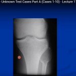
Case 15: Myxoid Liposarcoma
Clinical Features
- Large deep soft tissue mass near knee
Imaging
- Heterogeneous lesion deep to fascia
Histology
Findings include:
- Lipoblasts
- Myxoid stroma
- Delicate branching vasculature
Cytogenetics
Associated translocation:
- t(12;16)
Treatment
- Wide resection
- Radiotherapy
Case 16: Leiomyosarcoma
Clinical Features
- Deep thigh mass
- Mild exercise-related discomfort
Histology
- Spindle cells
- Corkscrew/cigar-shaped nuclei
Immunohistochemistry
Positive markers:
- Smooth muscle actin
- Desmin
Common Sites
- Pelvis
- Gastrointestinal tract
- Retroperitoneum
Treatment
- Wide excision
- Radiotherapy
Case 17: Clear Cell Sarcoma
Clinical Features
- Painful foot mass
- Usually near tendons
Histology
- Spindle cell sarcoma
- Positive melanocytic markers
Important Point
Also called:
- Melanoma of soft parts
Common Location
- Distal extremities
Treatment
- Wide resection
- Radiotherapy
Case 18: Parosteal Osteosarcoma
Clinical Features
- Slow-growing surface bone tumor
- Adult patient
Imaging Findings
- Heavily ossified cauliflower-like mass
- Attached to cortex
- Cleavage plane between tumor and cortex
Histology
- Low-grade fibroblastic tumor
- Bone production
Prognosis
- Favorable prognosis
Treatment
- Wide resection
- Chemotherapy rarely required
Case 19: Conventional Osteosarcoma
Clinical Features
- Young adult
- Aggressive proximal fibular lesion
Imaging Findings
- Permeative bone destruction
- Aggressive periosteal reaction
- Ossified matrix
Histology
- Malignant osteoid production
Standard Treatment
- Neoadjuvant chemotherapy
- Surgical resection
- Adjuvant chemotherapy
Case 20: Osteoid Osteoma
Clinical Features
Classic presentation:
- Young adult
- Severe nocturnal pain
- Pain relieved by NSAIDs
CT Findings
- Small nidus
- Surrounding sclerosis
MRI Findings
- Extensive marrow edema
Histology
- Woven bone trabeculae
- Osteoblast lining
- Vascular stroma
Treatment
- Radiofrequency ablation
Important Oncology Principles
Diagnosis Depends On
- Age of patient
- Tumor location
- Imaging characteristics
- Histology
- Immunohistochemistry
Imaging Clues
| Feature | Suggestive Diagnosis |
|---|---|
| Ring-and-arc calcification | Chondroid tumor |
| Dense ossification in soft tissue | Extraskeletal osteosarcoma |
| Target sign on MRI | Schwannoma |
| Triple signal intensity | Synovial sarcoma |
| Small nidus with sclerosis | Osteoid osteoma |
General Management Principles
- Biopsy before definitive treatment
- Wide oncologic margins for malignant tumors
- Limb salvage whenever feasible
- Multidisciplinary sarcoma care essential
- Reconstruction often required after resection
Key Exam Pearls
- Dedifferentiated chondrosarcoma has very poor prognosis
- PVNS contains hemosiderin-laden macrophages
- Schwannoma shows Antoni A/B areas and Verocay bodies
- Osteoid osteoma pain improves with NSAIDs
- Parosteal osteosarcoma is low grade and surface-based
- Conventional osteosarcoma requires chemotherapy plus surgery
Related Posts
 Oncology Case Studies and Quiz- 2
Courtesy: Prof James Wittig Orthopaedic Oncologist Sarcoma Surgeon www.tumorsurgery.org James Wittig books:- Unknown Musculoskeletal…
 Oncology Quiz and Case Studies- Part 3
Courtesy: James Wittig,Orthopaedic oncologist Unknown Musculoskeletal Tumor Cases: Diagnostic Approach and Key Lessons Overview This…
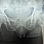 Image Quiz 14, #Evidence Case Study
Popular Answer may not imply right answer, Follow our twitter handle for the right answer,…
Leave a Reply