Courtesy: James Wittig
Orthopaedic Oncologist
Benign Soft Tissue Tumors: Structured Clinical Summary
Overview
- Benign soft tissue tumors arise from soft tissues such as skin, subcutaneous tissue, muscle, and connective tissue surrounding bone.
- They do not originate from bone and do not metastasize.
- Common entities include lipoma, hemangioma, fibromatosis, myxoma, schwannoma, neurofibroma, giant cell tumor of tendon sheath, and pigmented villonodular synovitis.
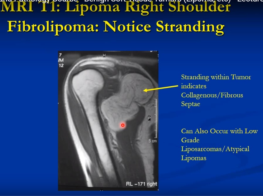
Lipoma
- Benign tumor composed of mature adipocytes with uniform nuclei and large cytoplasmic lipid vacuoles.
- Most common benign soft tissue tumor; commonly affects adults.
- Common sites include trunk, shoulder, neck, abdomen, and proximal extremities.
- Usually presents as a painless, slowly enlarging mass.
- MRI shows signal characteristics identical to subcutaneous fat on all sequences.
- Treatment is observation for asymptomatic lesions or marginal excision for symptomatic lesions.
- Recurrence is rare but higher in intramuscular lesions.
Hemangioma
- Benign proliferation of mature vascular channels, often congenital malformations rather than true neoplasms.
- May occur in skin, subcutaneous tissue, muscle, synovium, or bone.
- Intramuscular lesions often present with pain and enlargement after activity.
- Radiographs may show phleboliths; MRI shows heterogeneous lesion with serpiginous vascular channels and fat components.
- Treatment depends on symptoms and may include observation or surgical excision.
Fibromatosis
- Benign but locally aggressive fibroproliferative tumor with infiltrative growth pattern.
- Common in patients aged 15 to 40 and slightly more common in females.
- Common sites include shoulder girdle, upper arm, buttock, trunk, and head and neck.
- MRI shows infiltrative margins with variable signal depending on collagen content.
- Treatment is wide excision; recurrence rates are high.
- Radiotherapy and systemic therapy may be considered in selected cases.
Myxoma
- Rare benign hypocellular tumor composed of bland spindle and stellate fibroblasts in abundant myxoid stroma.
- Typically affects adults aged 40 to 60 years and commonly arises in thigh or upper arm.
- MRI shows homogeneous low signal on T1 and high signal on T2 due to mucin content.
- Treatment is marginal excision with low recurrence rate.
Schwannoma
- Benign nerve sheath tumor arising from Schwann cells.
- Usually affects adults aged 20 to 50 and commonly involves major peripheral nerves.
- Presents with pain and positive Tinel sign in many patients.
- MRI shows well circumscribed mass with split fat sign and target sign.
- Treatment is surgical excision with preservation of the nerve.
Neurofibroma
- Benign tumor of peripheral nerves, often solitary but associated with neurofibromatosis in some cases.
- Usually painless and located in superficial nerves.
- Unlike schwannoma, it infiltrates nerve fascicles and cannot be separated from the nerve.
- Surgical excision is rarely performed unless malignancy is suspected.
Giant Cell Tumor of Tendon Sheath
- Localized nodular tenosynovitis composed of synovial like cells, giant cells, inflammatory cells, and hemosiderin.
- Most common benign soft tissue tumor of the hand.
- MRI shows lesion with low signal areas due to hemosiderin deposition.
- Treatment is surgical excision; recurrence can occur.
Pigmented Villonodular Synovitis
- Diffuse or localized synovial proliferative disorder most commonly affecting the knee.
- Presents with joint swelling, pain, and recurrent effusion.
- MRI shows synovial mass with low signal from hemosiderin and blooming on gradient sequences.
- Treatment is synovectomy; recurrence rates are high in diffuse disease.

Leave a Reply