Courtesy: Prof James Wittig
Orthopaedic Oncologist
Sarcoma Surgeon
www.tumorsurgery.org
James Wittig books
Soft Tissue Sarcomas
Overview
Soft tissue sarcomas are malignant tumors arising from mesenchymal tissues such as:
- Muscle
- Fat
- Fibrous tissue
- Blood vessels
- Peripheral nerves
They may occur in:
- Extremities
- Pelvis
- Retroperitoneum
- Trunk
- Head and neck
Common histological types include:
- Liposarcoma
- Undifferentiated pleomorphic sarcoma
- Leiomyosarcoma
- Synovial sarcoma
- Fibrosarcoma
- Rhabdomyosarcoma
Most occur in adults between 40–70 years, except:
- Rhabdomyosarcoma: common in children
- Synovial sarcoma: common in adolescents and young adults
General Clinical Features
Typical presentation:
- Painless enlarging soft tissue mass
Important features concerning for malignancy:
- Size >5 cm
- Deep location beneath fascia
- Rapid growth
- Recurrence after previous excision
Pain is less common, although:
- Synovial sarcoma may be painful
Metastatic Pattern
Most common metastatic site:
- Lungs
Other sites:
- Bone
- Liver
Imaging Characteristics
MRI Findings
MRI is the investigation of choice.
Typical findings:
- Heterogeneous T1 and T2 signals
- Necrosis
- Hemorrhage
Special MRI Features
Myxoid Tumors
- Homogeneous appearance
- Hyperintense on T2
- Due to mucopolysaccharide content
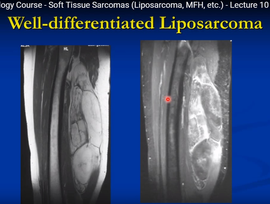
Well-Differentiated Liposarcoma
- Fat signal intensity
- Thick septations
- Nodular areas
Synovial Sarcoma
May show:
- Calcifications
- Triple signal intensity on T2 imaging
- Solid areas
- Hemorrhagic areas
- Cystic areas
Common Locations
| Tumor | Common Site |
|---|---|
| Liposarcoma | Thigh, retroperitoneum |
| Undifferentiated pleomorphic sarcoma | Thigh |
| Fibrosarcoma | Deep soft tissues of extremities |
| Synovial sarcoma | Near large joints |
| Epithelioid sarcoma | Hand |
| Leiomyosarcoma | Retroperitoneum |
Important Cytogenetic Translocations
Synovial Sarcoma
- t(X;18)
- Produces SYT-SSX fusion gene
Myxoid / Round Cell Liposarcoma
- t(12;16)
Alveolar Rhabdomyosarcoma
- t(2;13) or t(1;13)
Immunohistochemistry
Most sarcomas are:
- Vimentin positive
Additional markers help identify lineage.
Examples:
| Tumor | Marker |
|---|---|
| Leiomyosarcoma | Actin, desmin |
| Rhabdomyosarcoma | Myogenin, desmin |
| Synovial sarcoma | Cytokeratin, EMA |
Liposarcoma
Overview
- Second most common soft tissue sarcoma
- Most common sarcoma in adults
Subtypes
- Well differentiated
- Myxoid
- Round cell
- Pleomorphic
- Dedifferentiated
Histology
Well Differentiated
- Resembles lipoma
- Lipoblasts present
- Thick atypical septa
Myxoid
- Myxoid matrix
- Arborizing capillaries
Treatment
- Wide surgical excision
- Radiotherapy depending on margins and location
Undifferentiated Pleomorphic Sarcoma
Previously called:
- Malignant fibrous histiocytoma
Features
- High-grade sarcoma
- Pleomorphic spindle cells
- Deep soft tissue location
- Common in extremities and retroperitoneum
Histology
- Storiform pattern
- Marked pleomorphism
- Necrosis common
Treatment
- Wide excision
- Radiotherapy
Fibrosarcoma
Features
- Malignant fibroblast tumor
- Usually deep soft tissue lesion
Histology
Classic:
- Herringbone pattern
Treatment
- Wide excision
- Radiotherapy
Synovial Sarcoma
Overview
- Common near joints
- Rarely intra-articular
- Typically affects young adults
Histological Types
Biphasic
- Spindle cells
- Epithelial cells
Monophasic
- Mainly spindle cells
Imaging Features
- Calcification
- Cystic degeneration
- Triple signal intensity on MRI
Treatment
- Wide excision
- Radiotherapy
- Chemotherapy often used in younger patients
Rhabdomyosarcoma
Overview
- Most common malignant soft tissue tumor in children
Subtypes
- Embryonal
- Alveolar
- Pleomorphic
Histology
- Rhabdomyoblasts
- Cross striations may be seen
Treatment
Multimodal treatment:
- Surgery
- Chemotherapy
- Radiotherapy
Leiomyosarcoma
Overview
- Malignant smooth muscle tumor
Common sites:
- Retroperitoneum
- Deep soft tissues
Histology
- Spindle cells
- Cigar-shaped nuclei
- Actin and desmin positive
Treatment
- Wide surgical excision
- Radiotherapy
Management Principles
Mainstay of Treatment
Wide Surgical Excision
Goals:
- Adequate margins
- Limb preservation
- Local control
Radiotherapy
Used for:
- Local control
- High-grade tumors
- Large tumors
- Close margins
Chemotherapy
Selective use in:
- Rhabdomyosarcoma
- Synovial sarcoma
- Metastatic disease
Amputation
Reserved for:
- Unresectable tumors
- Extensive contamination
- Nonfunctional limb
Prognostic Factors
Poor prognostic indicators:
- Large tumor size
- High grade
- Deep location
- Metastases
- Positive surgical margins
Prognosis
- Five-year survival for nonmetastatic disease: approximately 60–65%
- Metastatic disease significantly worsens prognosis
Outcome depends on:
- Histological grade
- Tumor size
- Tumor location
- Completeness of surgical resection
Related Posts
 Principles of treating Soft tissue Sarcomas
Courtesy: Prof Robert Ashford, President, British Orthopaedic Oncology Society Principles of Treating Soft Tissue…
- Principles of treating Soft tissue Sarcomas
Courtesy: Prof Robert Ashford, President, British Orthopaedic Oncology Society Principles of Treating Soft Tissue…
 Benign Soft Tissue Tumours
Courtesy: James Wittig Orthopaedic Oncologist Benign Soft Tissue Tumors: Structured Clinical Summary Overview Benign soft…
Leave a Reply