Courtesy: Derek Lohan MD David Geffen School of Medicine UCLA 10833 Le Conte Ave STE 428 Los Angeles, CA 90095
-Applied Anatomy
Osteoclast function and Roles

? Courtesy: Prof Nabil Ebraheim, University of Toledo, Ohio, USA Osteoclast is a large multinucleated cell. It has 15- 20 nuclei. Its origin is from the fusion of macrophages /monocytes. As many as 50 of cells fuse to form a giant osteoclast. Osteoclast absorbs bone. Its function is regulated by osteoblast. Life span is of […]
Anatomy and Innervation of the Lumbar Plexus
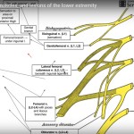
Courtesy: Medical Lectures Made Easy
Acetabular fractures: Applied Anatomy
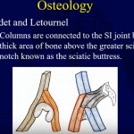
Courtesy: Saqib Rehman MD Director of Orthopaedic Trauma Temple University Philadelphia Pennsylvania USA
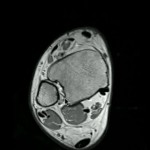
Types of Joints: Anatomic and Radiologic Correlation
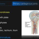
Courtesy: Andrew Dixon MD Radiologist Alfred Health Melbourne, Australia and Radiopedia www.radiopaedia.org
Sciatica Anatomy
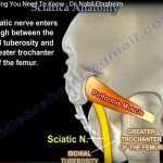
Courtesy: Prof Nabil Ebraheim University of Toledo, Ohio, USA Anatomy of sciatica The sciatic nerve is a large nerve that originates from the lumbosacral plexus of lower spine with a root value of L4, L5, S1, S2& S3 . The nerve initially emerges from the pelvis and leaves the greater sciatic notch, anterior and deep […]
The Superior Gluteal Artery
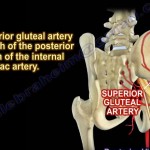
Courtesy: Prof Nabil Ebraheim, University of Toledo, Ohio, USA
Anatomy of Coracoid, Conoid and Coronoid
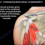
Courtesy: Prof Nabil Ebraheim, University of Toledo, Ohio, USA This video speaks about the coracoid, conoid, coronoid processes. Coracoid is the hook shaped bony process of scapula. It is considered as the surgeons lighthouse. It is safe to do surgical approaches lateral to coracoid process. The neuro vascular bundle lies medial to coracoid process. Three […]
Anatomy of Brachialis, Coracobrachialis and Brachioradialis
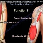
Courtesy: Prof Nabil Ebraheim, University of Toledo, Ohio, USA
Anatomy of Gracilis Muscle

Courtesy: Prof Nabil Ebraheim, University of Toledo, Ohio, USA GRACILIS MUSCLE ANATOMY It is the most superficial muscle on the medial side of thigh The tendon of gracilis muscle is easily palpable in the inguinal region together with adductor longus muscle. Medial side of the thigh contains the adductor group of muscles which help […]
Gluteus Medius #Anatomy
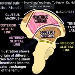
Courtesy: Prof Nabil Ebraheim, University of Toledo, Ohio, USA This video describes anatomy of gluteus medius muscle. It originates from dorsal ilium inferior to iliac crest from posterior gluteal line to anterior gluteal line and inserts into lateral and superior aspect of greater trochanter of femur. Gluteus medius forms the middle layer of gluteal muscles, […]
Acromioclavicular Joint #Anatomy and Classification of Injury
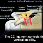
Courtesy: Prof Nabil Ebraheim, University of Toledo, Ohio, USA
Extensor Muscles of #thumb
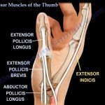
Courtesy: Prof Nabil Ebraheim, University of Toledo, Ohio, USA EXTENSOR MUSCLES OF THUMB Extensor Pollicis Brevis Extensor Pollicis Longus Abductor Pollicis Longus Extensor Pollicis Brevis Origin: posterior surface of Radius and interosseous membrane Insertion: Proximal phalanx of thumb Extensor Pollicis Longus Origin: posterior surface of Ulna and interosseous membrane Insertion: Distal phalanx of thumb Abductor […]
Anatomy of #Extensor Pollicis Brevis
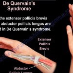
Courtesy: Prof Nabil Ebraaheim, University of Toledo, Ohio, USA Origin Posterior surface of the radius Interosseous membrane of the forearm Insertion Base of the proximal phalanx of the thumb Nerve Supply Posterior interosseous nerve Function Extension of the thumb at the metacarpophalangeal joint Anatomical Relationships Forms the lateral border of the anatomical snuffbox Lies in […]
Anatomy of Abductor #Pollicis Longus
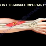
Courtesy: Prof Nabil Ebraheim, University of Toledo, Ohio, USA ABDUCTOR POLLICIS LONGUS • The Abductor Pollicis Longus is an important muscle . • It is one of the extrinsic muscles of the hand. ORIGIN: It arises from the posterior surface of the ulna, radius and the interosseous membrane. INSERTION: The APL is inserted into the […]