Courtesy: Derek Lohan MD
David Geffen School of Medicine
MRI Anatomy of the Ankle
Introduction
MRI of the ankle is an essential imaging modality used to evaluate:
- Tendons
- Ligaments
- Bones
- Joint structures
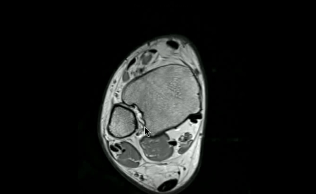
Tendon Compartments of the Ankle
Tendons around the ankle are grouped into three compartments:
- Flexor compartment (posteromedial)
- Peroneal compartment (lateral)
- Extensor compartment (anterior)
Important Ligament Groups
- Syndesmotic ligaments
- Lateral ligament complex
- Deltoid ligament
- Spring ligament
Tendons Around the Ankle
1. Flexor Compartment (Posteromedial)
Mnemonic: Tom, Dick And Harry
| Structure | Meaning |
|---|---|
| T | Tibialis posterior |
| D | Flexor digitorum longus |
| A | Posterior tibial artery |
| N | Tibial nerve |
| H | Flexor hallucis longus |
Key Point
- Located behind the medial malleolus
Flexor Hallucis Longus
- Easily identified on MRI because:
- Muscle belly extends more distally than other flexors
Achilles Tendon
Location
- Posterior aspect of ankle
Function
- Plantarflexion
Formation
- Gastrocnemius + Soleus tendons
2. Peroneal Compartment (Lateral)
Tendons
- Peroneus longus
- Peroneus brevis
Identification Tip
- Brevis = Bone
- Lies closer to fibula
- Longus lies more superficial
3. Extensor Compartment (Anterior)
Mnemonic: Tom, Harry, Dick
| Structure | Meaning |
|---|---|
| T | Tibialis anterior |
| H | Extensor hallucis longus |
| D | Extensor digitorum longus |
Location
- Anterior to ankle joint
Distal Tibiofibular Syndesmosis
Definition
- Fibrous joint between distal tibia and fibula
Ligaments
- Anterior tibiofibular ligament
- Posterior tibiofibular ligament
Function
- Stabilizes ankle mortise
Clinical Importance
- Injured in high ankle sprains
Lateral Ligament Complex
1. Anterior Talofibular Ligament (ATFL)
Features
- Fibula – Talus
- Thin and horizontal
- Most commonly injured ligament
Mechanism of Injury
- Inversion injury
2. Posterior Talofibular Ligament (PTFL)
Features
- Strong and thick
- Rarely injured
3. Calcaneofibular Ligament (CFL)
Features
- Fibula – Calcaneus
- Vertical orientation
- Lies deep to peroneal tendons
Deltoid Ligament (Medial Side)
Characteristics
- Strong triangular ligament
- Stronger than lateral ligaments
Components
- Superficial
- Deep
Function
- Provides medial ankle stability
Clinical Importance
- Injury requires significant trauma
Spring Ligament (Calcaneonavicular Ligament)
Attachment
- Calcaneus — Navicular
Functions
- Supports talar head
- Maintains medial longitudinal arch
Clinical Importance
- Injury leads to:
- Flatfoot deformity
- Posterior tibial tendon dysfunction
Sinus Tarsi
Location
- Between talus and calcaneus
Contents
- Ligaments
- Fat
Clinical Importance
- Inflammation — Sinus tarsi syndrome
MRI Imaging Planes
Axial Plane
- Best for:
- Tendons
- Lateral ligaments
- Syndesmotic ligaments
Coronal Plane
- Best for:
- Deltoid ligament
- Calcaneofibular ligament
- Joint alignment
Sagittal Plane
- Best for:
- Achilles tendon
- Plantar fascia
- Talocalcaneal ligaments
Plantar Fascia
Location
- Plantar aspect of foot
Functions
- Supports longitudinal arch
- Absorbs shock
Clinical Condition
- Plantar fasciitis — common cause of heel pain
Summary
Tendon Compartments
- Flexor (posteromedial)
- Peroneal (lateral)
- Extensor (anterior)
Important Ligaments
Lateral
- ATFL
- PTFL
- CFL
Medial
- Deltoid ligament
Others
- Syndesmotic ligaments
- Spring ligament
Key Clinical Points
- ATFL = most commonly injured ligament
- Deltoid ligament injuries are rare
- CFL lies deep to peroneal tendons
- Spring ligament supports medial arch

Leave a Reply