Courtesy: Dr Jean Jose MD, Associate Chief, Musculoskeletal Radiology Section, Associate Professor of Clinical Radiology, University of Miami School of Medicine, Florida, USA
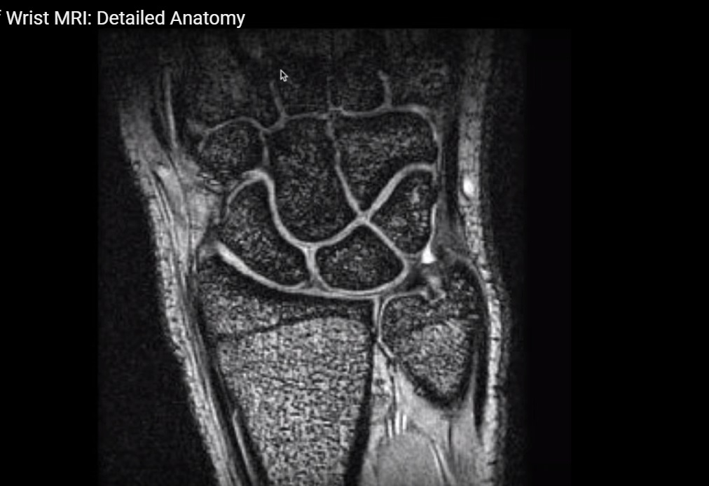
MRI Anatomy of the Wrist
Overview
The wrist is a complex anatomical region consisting of:
- Multiple bones
- Tendons
- Ligaments
- Neurovascular structures
Clinical Importance
MRI of the wrist is essential for evaluating:
- Ligament injuries
- Tendon pathology
- Carpal instability
Main Components
- Extensor tendons (dorsal)
- Flexor tendons (volar/palmar)
- Carpal bones
- Intrinsic and extrinsic ligaments
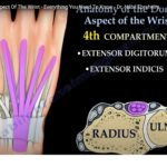
Dorsal (Extensor) Tendon Compartments
Key Landmark
- Lister’s tubercle (distal radius)
Six Dorsal Compartments
| Compartment | Tendons |
|---|---|
| 1st | Abductor pollicis longus (APL), Extensor pollicis brevis (EPB) |
| 2nd | Extensor carpi radialis longus (ECRL), Extensor carpi radialis brevis (ECRB) |
| 3rd | Extensor pollicis longus (EPL) |
| 4th | Extensor digitorum communis (EDC), Extensor indicis |
| 5th | Extensor digiti minimi |
| 6th | Extensor carpi ulnaris (ECU) |
Clinical Point
- EPL crosses over:
- Lister’s tubercle
- Second compartment tendons
Can lead to Intersection Syndrome
Volar (Flexor) Tendons
Carpal Tunnel Contents
Total: 9 Tendons
- Flexor digitorum profundus (4)
- Flexor digitorum superficialis (4)
- Flexor pollicis longus (1)
Nerve in Carpal Tunnel
- Median nerve
Position
- Lies between:
- FDS and FDP tendons
Important Note
- Flexor carpi radialis:
- Runs in a separate canal
- Not part of carpal tunnel
Order (Lateral — Medial)
- Flexor carpi radialis
- Flexor pollicis longus
- Flexor digitorum superficialis
- Flexor digitorum profundus
- Flexor carpi ulnaris
Important Nerves of the Wrist
Median Nerve
- Passes through carpal tunnel
- Supplies most flexor muscles
Ulnar Nerve
- Passes through Guyon’s canal
- Located near pisiform
Radial Nerve (Superficial Branch)
- Near radial styloid
- Supplies sensation to:
- Thumb
- Index finger
Important Arteries
Radial Artery
- Runs along volar radial side
- Passes deep to first dorsal compartment
Ulnar Artery
- Travels with ulnar nerve in:
- Guyon’s canal
Carpal Bones
Total: 8 Bones
Proximal Row
- Scaphoid
- Lunate
- Triquetrum
- Pisiform
Distal Row
- Trapezium
- Trapezoid
- Capitate
- Hamate
Hook of Hamate
- Clinically important in:
- Ulnar nerve compression
- Sports injuries
Wrist Joints
Radiocarpal Joint
- Radius + Scaphoid + Lunate
Distal Radioulnar Joint (DRUJ)
- Radius + Ulna
Function
- Pronation and supination
Ligaments of the Wrist
1. Intrinsic Ligaments
- Between carpal bones
Examples
- Scapholunate ligament
- Lunotriquetral ligament
2. Extrinsic Ligaments
- Connect carpal bones to radius/ulna
Examples
- Radioscaphocapitate ligament
- Radiolunate ligament
- Ulnolunate ligament
Scapholunate Ligament
Components
- Dorsal
- Volar
- Proximal fibrocartilage
Most Important
- Dorsal component
Function
- Provides wrist stability
Clinical Importance
- Injury leads to:
- Scapholunate dissociation
- Carpal instability
Lunotriquetral Ligament
Components
- Dorsal
- Volar
- Proximal
Function
- Stabilizes lunate–triquetrum joint
Volar Extrinsic Ligaments
Important Ligaments
- Radioscaphocapitate ligament
- Long radiolunate ligament
- Short radiolunate ligament
- Ulnolunate ligament
- Ulnotriquetral ligament
Radioscaphocapitate Ligament
- Extends:
- Radius — Scaphoid — Capitate
- Part of arcuate ligament complex
Long Radiolunate Ligament
- Also called:
- Radiolunotriquetral ligament
Dorsal Wrist Ligaments
Main Ligaments
- Dorsal intercarpal ligament
- Dorsal radiotriquetral ligament
- Dorsal distal radioulnar ligament
Dorsal Intercarpal Ligament
- Extends:
- Triquetrum — Scaphoid
- Also attaches to trapezium & trapezoid
First Carpometacarpal (CMC) Joint Ligaments
Important for Thumb Stability
- Anterior oblique ligament
- Dorsal radial ligament
- Deep anterior oblique ligament
Clinical Correlations
1. Carpal Tunnel Syndrome
- Compression of:
- Median nerve
Symptoms
- Numbness
- Tingling
- Thenar weakness
2. Intersection Syndrome
- Occurs at:
- EPL crossing second compartment
Symptoms
- Pain over dorsal wrist
3. Scapholunate Ligament Injury
- Leads to:
- Carpal instability
Common Cause
- FOOSH (fall on outstretched hand)
Summary
Key Structures
- Six dorsal extensor compartments
- Nine flexor tendons in carpal tunnel
- Eight carpal bones
- Intrinsic and extrinsic ligaments
- Major nerves:
- Median
- Ulnar
- Radial
Clinical Importance
- Essential for diagnosing:
- Tendon injuries
- Ligament tears
- Carpal instability

Leave a Reply