Courtesy: Medical Lectures Made Easy
Introduction
The lumbar plexus is a complex network of nerves formed by the ventral rami of spinal nerves L1 to L4, with occasional contribution from T12.
Location
The plexus is located within the psoas major muscle, situated along the posterior abdominal wall.
Functions
The lumbar plexus provides motor and sensory innervation to:
-
Lower abdominal wall
-
Anterior thigh
-
Medial thigh
-
Genital region (cutaneous supply)
-
Portions of the lower limb
Branches of the Lumbar Plexus
The major branches include:
-
Ilioinguinal nerve
-
Genitofemoral nerve
-
Lateral femoral cutaneous nerve
-
Femoral nerve
-
Obturator nerve
1. Ilioinguinal Nerve
Root
-
L1
Course
-
Travels through the inguinal canal
Function
Provides sensory innervation to:
-
Scrotum (male)
-
Labia majora (female)
-
Upper medial thigh
2. Genitofemoral Nerve
Roots
-
L1–L2
Divisions
The nerve divides into:
-
Genital branch
-
Femoral branch
Genital Branch
Course
-
Passes through the inguinal canal
Function
-
Sensory supply to scrotum or labia majora
Femoral Branch
Course
-
Travels beneath the inguinal ligament
Function
-
Supplies skin over the proximal anterior thigh
3. Lateral Femoral Cutaneous Nerve
Roots
-
L2–L3
Course
-
Passes beneath the inguinal ligament
Function
-
Provides sensory innervation to the anterolateral thigh
Clinical Correlation: Meralgia Paresthetica
Cause
-
Compression of the lateral femoral cutaneous nerve
Symptoms
-
Burning pain
-
Tingling sensation
-
Numbness over the anterolateral thigh
Common Risk Factors
-
Tight clothing
-
Obesity
-
Pregnancy
-
Pelvic compression
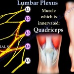
4. Femoral Nerve
Roots
-
L2–L4
Course
-
Emerges from the lateral border of psoas major
-
Passes beneath the inguinal ligament
-
Enters the femoral triangle
Distribution
-
Supplies muscles of the anterior thigh
Muscles Supplied by the Femoral Nerve
Hip Flexors
-
Pectineus
-
Sartorius
-
Iliopsoas
Iliopsoas Muscle
Function
-
Primary hip flexor
-
Assists in lateral rotation
Insertion
-
Lesser trochanter of femur
Knee Extensors (Quadriceps Femoris)
Muscles include:
-
Rectus femoris
-
Vastus medialis
-
Vastus lateralis
-
Vastus intermedius
Function
-
Extension of the knee
Insertion
-
Patella – Patellar tendon andTibial tuberosity
Saphenous Nerve
Key Features
-
Terminal branch of femoral nerve
-
Longest sensory nerve in the body
-
Passes through the adductor canal
Sensory Supply
-
Medial leg
-
Medial aspect of foot
Adductor Canal
Contents
-
Femoral artery
-
Femoral vein
-
Saphenous nerve
-
Nerve to vastus medialis
Clinical Correlation: Femoral Neck Fracture
Presentation
-
Shortened limb
-
External rotation
Mechanism
The iliopsoas muscle pulls the limb into:
-
Flexion
-
External rotation
Additional Muscles
Sartorius
-
Functions: Hip flexion, abduction, lateral rotation
-
Innervation: Femoral nerve
Pectineus
-
Functions: Hip flexion and adduction
-
Innervation:
-
Primarily femoral nerve
-
Occasionally obturator nerve
-
5. Obturator Nerve
Roots
-
L2–L4
Course
-
Passes through the obturator canal
Distribution
-
Supplies the medial compartment of the thigh
Muscles Supplied
-
Adductor longus
-
Adductor brevis
-
Adductor magnus (partial)
-
Gracilis
-
Obturator externus
Function
-
Primary action: Adduction of the thigh
Gracilis Muscle
Features
-
Long, slender muscle
-
Commonly used in graft procedures
Clinical Note
-
Functionally expendable
Pes Anserinus
Components
Insertion of three muscles:
-
Sartorius
-
Gracilis
-
Semitendinosus
Location
-
Medial surface of the proximal tibia
Meaning
-
Latin for “Goose Foot”
Adductor Hiatus
Description
An opening in the adductor magnus muscle
Structures Passing Through
-
Femoral artery
-
Femoral vein
Continuation
These vessels become:
-
Popliteal artery
-
Popliteal vein
Connection to Sacral Plexus
Lumbosacral Trunk
-
Formed by: L4 and L5 nerve roots
-
Connects lumbar plexus to sacral plexus
Summary Table
| Nerve | Roots | Function |
|---|---|---|
| Ilioinguinal | L1 | Sensory to genital region & upper thigh |
| Genitofemoral | L1–L2 | Sensory to genital region & proximal thigh |
| Lateral femoral cutaneous | L2–L3 | Sensory to lateral thigh |
| Femoral | L2–L4 | Motor to anterior thigh |
| Obturator | L2–L4 | Motor to medial thigh |
Key Clinical Conditions
-
Meralgia paresthetica ? Compression of lateral femoral cutaneous nerve
-
Femoral neck fracture ? Shortened, externally rotated limb
-
Adductor muscle injuries ? May involve obturator nerve

Leave a Reply