Courtesy: Saqib Rehman MD, Director of Orthopaedic Trauma
Introduction
Understanding acetabular fractures requires a solid grasp of:
- Acetabular anatomy
- Column concept
- Vascular anatomy
- Neural anatomy
- Radiographic interpretation
Historical Background
Modern classification is based on the work of:
- Robert Judet
- Émile Letournel
Their Judet–Letournel classification remains the gold standard
Osteology of the Acetabulum
Components
The acetabulum is formed by the fusion of:
- Ilium
- Ischium
- Pubis
Articular Surface
- Shaped like an inverted horseshoe
- Surrounds the acetabular fossa
Key Structural Elements
- Anterior column
- Posterior column
- Acetabular roof (weight-bearing dome)
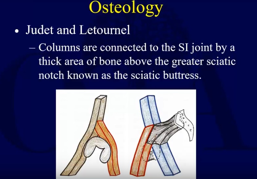
Column Concept of the Acetabulum
Inverted Y Model
The acetabulum can be visualized as an inverted Y structure:
- One limb – Anterior column
- Other limb – Posterior column
The articular surface lies between these two columns
Anterior Column
Also Called
- Iliopubic column
Extent
- Iliac crest – acetabulum – pubic symphysis
Components
- Iliac segment
- Acetabular segment
- Pubic segment
Posterior Column
Extent
- Greater sciatic notch – posterior acetabulum – ischial tuberosity
Function
- Forms the posterior wall of the acetabulum
Sciatic Buttress
Location
- Above the greater sciatic notch
Importance
- Connects:
- Anterior column
- Posterior column
- Sacroiliac joint
Major load-transmitting structure
Acetabular Roof (Weight-Bearing Dome)
Definition
- Superior portion of acetabulum
Function
- Transfers load from femoral head to pelvis
Clinical Importance
Fractures involving this area are highly significant
3D Orientation of the Acetabulum
Direction
- Faces:
- Laterally
- Inferiorly
- Anteriorly
Important Vascular Anatomy
Obturator Artery
Origin
- Internal iliac artery
Function
- Supplies pelvic structures and hip joint
Corona Mortis (Crown of Death)
Definition
- Vascular connection between:
- Obturator artery
- Inferior epigastric / external iliac artery
Location
- Retropubic region
- Superior pubic ramus
Clinical Importance
Injury leads to:
- Severe hemorrhage
- Difficult surgical control
Blood Supply of Femoral Head
Primary Source
- Medial femoral circumflex artery (MFCA)
Course
- Deep to quadratus femoris
- Near obturator internus
- Posterior to femoral neck
Clinical Importance
Injury can cause:
- Avascular necrosis (AVN)
Surgical Tip
- Preserve quadratus femoris
- Leave muscle tags on short external rotators
Superior Gluteal Artery
Course
- Through greater sciatic notch
- Above piriformis
Risk
- Injury during posterior approaches
- Aggressive retraction
Neural Anatomy Around the Acetabulum
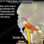
Sciatic Nerve
Significance
- Most commonly injured nerve
Causes
- Trauma
- Posterior hip dislocation
- Surgery
Prevention
- Hip extension
- Knee flexion
Reduces nerve tension
Complication
- Foot drop
Superior Gluteal Nerve
Function
- Supplies:
- Gluteus medius
- Gluteus minimus
Injury Result
- Trendelenburg gait
Inferior Gluteal Nerve
Function
- Supplies gluteus maximus
Surgical Risk
- Avoid high splitting of gluteus maximus
Greater Sciatic Notch: Key Zone
Structures Passing Through
- Sciatic nerve
- Superior gluteal nerve
- Superior gluteal artery
- Inferior gluteal nerve
Clinical Importance
High-risk area during:
- Retraction
- Surgical exposure
Summary
Acetabular Anatomy
- Two columns:
- Anterior
- Posterior
- Roof = weight-bearing dome
Important Vessels
- Obturator artery
- Corona mortis
- Medial femoral circumflex artery
- Superior gluteal artery
Important Nerves
- Sciatic nerve
- Superior gluteal nerve
- Inferior gluteal nerve
Key Clinical Risks
- Sciatic nerve injury– foot drop
- Corona mortis injury– massive bleeding
- MFCA injury– AVN of femoral head
Key Take-Home Messages
- Think in columns (anterior + posterior)
- Protect vascular structures, especially corona mortis
- Always consider nerve safety during exposure
- Acetabular dome involvement = critical injury

Felicitaciones
Muchas gracias