

Courtesy: Sarav Shah, MD, Attending Orthopaedic Surgeon, New England Baptist Hospital, Boston, MA, USA Visit Dr Hitesh Gopalan
Knee Arthroplasty MCQs for the FRCS Orth

Courtesy: Zaid Al-Rab, Founder, OrthoPass, Leeds, UK Overview Catastrophic polyethylene wear is a serious complication in total knee replacement, often leading to implant failure. Contributing Factors 1. Surgical Factors Component malalignment Instability Poor ligament balancing 2. Patient Factors High activity levels Obesity 3. Polyethylene Factors Thin inserts Sterilization method Implant design Critical Threshold Polyethylene thickness […]
MRI Hip Made Easy
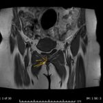
Courtesy: Syed Intekhab Alam, MSK Radiologist, Doha, Qatar
Patellar Resurfacing in TKR

Courtesy: Michael Decker, Assistant Professor and Arthroplasty Surgeon, University of New Mexico, ALbuquerque, USA
Remplissage and Labral Repair

Courtesy: Patrick Denard, MD, Oregon Shoulder Institute, Oregon, USA
Piriformis Syndrome
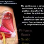
Courtesy: Prof Nabil Ebraheim, University of Toledo, Ohio, USA
Vitamin D Resistant Rickets

Courtesy: Prof Nabil Ebraheim, University of Toledo, Ohio, USA
Maisonneuve Fracture of Ankle
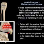
Courtesy: Prof Nabil EBraheim, University of Toledo, Ohio, USA Definition Maisonneuve fracture is a proximal fibular fracture associated with an unstable syndesmotic injury of the ankle. It is an important injury because the ankle injury may appear minor while significant instability exists at the syndesmosis. Why is it Commonly Missed? The injury may present without: […]
Update in the Management of Acetabular Defects
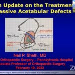
Courtesy: Neil Sheth, MD, Chief of Orthopaedcis, Hospital for the University of Pennsylvania, USA
Multi-Ligament Knee Injuries

Courtesy: Dr Peter MacDonald, FRCS, President-Elect, American Shoulder and Elbow Society, Past President, Canadian Orthopaedic Association
Coxa Vara in Children
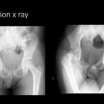
Courtesy: Prof Hitesh Shah, Professor of Paediatric Orthopaedics, KMC Manipal, India Coxa Vara in Children Introduction Coxa vara is a pediatric hip deformity characterized by a decreased femoral neck-shaft angle. The condition alters: Hip biomechanics Abductor function Limb alignment If progressive, it can lead to gait abnormalities, limb shortening, and long-term hip dysfunction. Normal […]
Nonunion of the Humerus
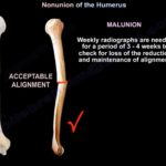
Courtesy: Prof Nabil Ebraheim, University of Toledo, Ohio, USA
Fracture Humerus and Radial Nerve Palsy
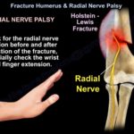
Courtesy: Prof Nabil Ebraheim, University of Toledo, Ohio, USA Radial nerve can be identified as it runs through the spiral groove between the heads of triceps. The nerve lies posterior to the deltoid tuberosity. The nerve crosses the posterior aspect of the humerus about 20 cm proximal to the medial epicondyle and about 15cm proximal […]
Elbow Pain: Causes and Treatment
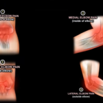
Courtesy: Prof Nabil Ebraheim, University of Toledo, Ohio, USA Introduction Elbow pain is best approached anatomically. Localizing the pain to a specific region of the elbow often helps narrow the diagnosis and guide treatment. The elbow can be divided into four regions: Anterior (front) Medial (inner side) Posterior (back) Lateral (outer side) This simple […]
Must-Know MCQs for the Fellowship Exam

Courtesy: Zaid Al-RUb, FRCS Orth, Founder, OrthoPass Courtesy: Zaid Al-Rub, Founder and CEO, OrthoPass Gap Balancing in Total Knee Replacement Scenario Primary Total Knee Replacement (TKR). After distal femur and proximal tibial cuts: Extension gap – tight Flexion gap – balanced First Step Posterior capsule release Principles of Gap Balancing Rule 1: Address soft tissue before additional […]