Courtesy: Prof Nabil Ebraheim, University of Toledo, Ohio, USA
Plantaris Tendon Rupture (Tennis Leg): Diagnosis and Management
Overview
Plantaris tendon rupture is a relatively uncommon but important cause of acute calf pain, often referred to as “tennis leg.”
It typically presents with a sudden onset of pain in the calf, frequently mimicking more serious conditions such as an Achilles tendon rupture.
Definition and Mechanism of Injury
Definition
- Rupture of the plantaris tendon
- Commonly associated with acute calf pain during activity
Mechanism
- Occurs due to:
- Eccentric loading of the ankle
- Knee in extended position
Typical Scenario
- Sudden push-off movement (e.g., tennis, running)
- Patient may describe:
- A “popping” sensation
- Feeling as if struck from behind
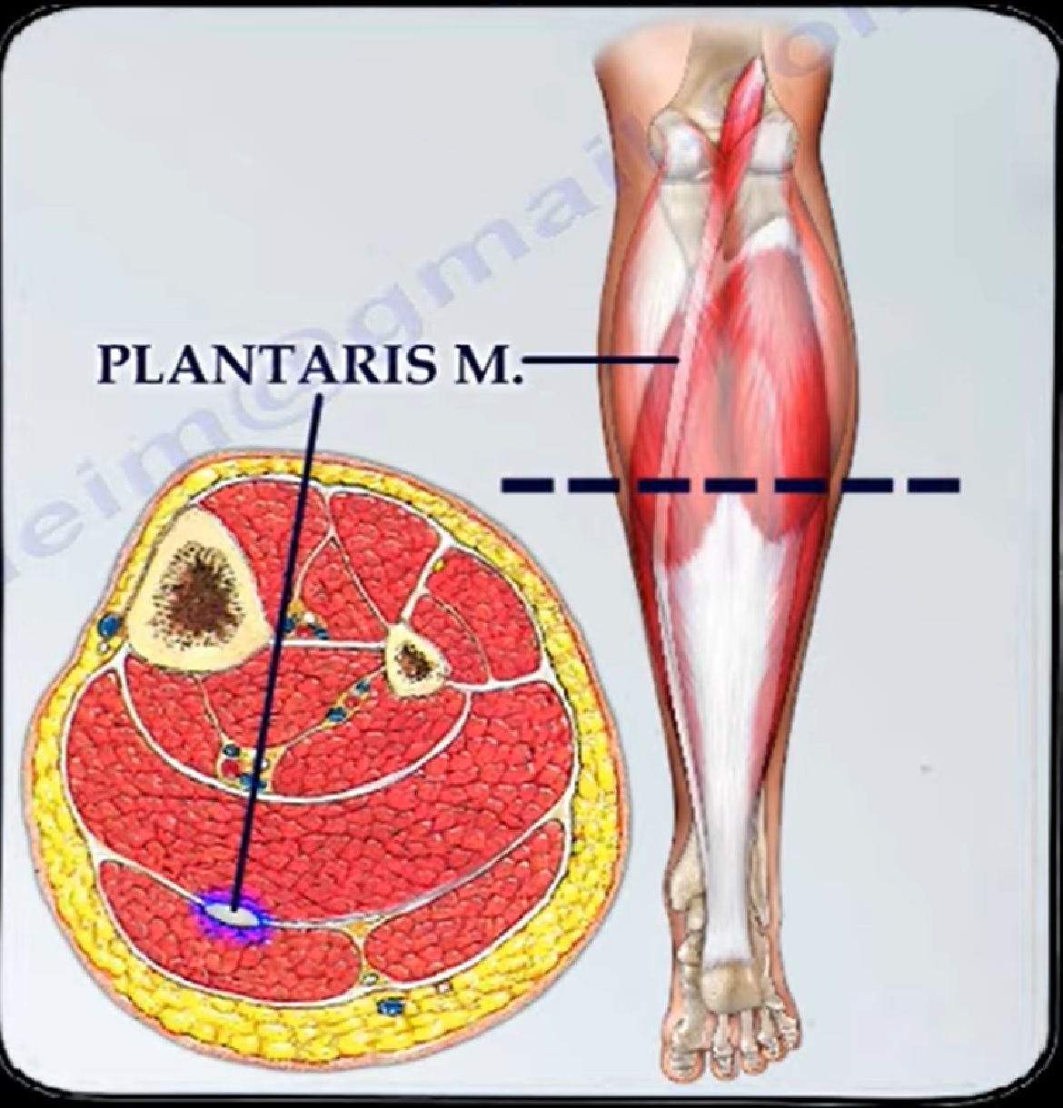
Anatomy of the Plantaris Muscle
Origin
- Lateral supracondylar ridge of femur
- Above lateral head of gastrocnemius
Insertion
- Medial aspect of the calcaneus
Location
- Superficial posterior compartment
- Lateral part of popliteal fossa
Function
- Weak plantarflexion of the ankle
- Assists in knee flexion
Innervation
- Tibial nerve
Additional Feature
- Rich in proprioceptive receptors
Contributes to positional feedback of the foot
Clinical Presentation
Symptoms
- Sudden sharp pain in the calf
- Sensation of tearing or snapping
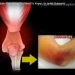
- Swelling of the calf
- Bruising (ecchymosis)
Functional Impact
- Walking: usually possible
- Running: significantly limited
Clinical Examination
Key Findings
- Tenderness in the calf
- Swelling and bruising
Important Consideration
- Can mimic Achilles tendon rupture
Thompson Test Interpretation
| Scenario | Result |
|---|---|
| Achilles + Plantaris rupture | Positive test |
| Achilles rupture with intact plantaris | May appear negative |
Careful interpretation is essential
Differential Diagnosis
- Achilles tendon rupture
- Gastrocnemius strain
- Deep vein thrombosis (DVT)
- Soleus muscle injury
Imaging
MRI Findings
T1-Weighted Images
- Loss of normal plantaris tendon visualization
T2-Weighted Images
- Edema in the surrounding tissues
Important Feature
- Achilles tendon remains intact (no discontinuity)
Management
Conservative Treatment (Preferred)
Initial Management
- Limb elevation
- Pain control
- Use of crutches if required
Immobilization
- CAM walker boot
Weight Bearing
- As tolerated
Prognosis
- Excellent recovery with non-operative treatment
- Most patients return to normal function
Additional Clinical Notes
- Plantaris tendon may be:
- Used as a graft in Achilles tendon reconstruction
- Injury may occur:
- In isolation
- Along with Achilles tendon rupture
Key Takeaways
- Plantaris rupture is a common mimic of Achilles tendon injury
- Sudden calf pain with a “pop” is characteristic
- Always rule out:
- Achilles rupture
- DVT
Management Summary
- Primarily conservative treatment
- Excellent outcomes expected
Clinical Insight
Accurate diagnosis is essential to:
- Avoid unnecessary surgical intervention
- Ensure appropriate rehabilitation
If you want, I can next:
- Create a quick comparison table: Plantaris rupture vs Achilles rupture vs Gastrocnemius tear
- Or a clinical diagnostic algorithm for acute calf pain
Leave a Reply