
Courtesy: Prof Nabil Ebraheim, University of Toledo, Ohio, USA
Quadriceps Tendon Rupture
1. Basic Concept
The quadriceps muscle is the large muscle group at the front of the thigh and is responsible for knee extension.
A quadriceps tendon rupture causes disruption of the extensor mechanism.
According to the lecture:
- Usually occurs in patients older than 40 years
- More common in males
- Usually occurs at the tendon insertion into the patella
- Typically just proximal to the superior pole of the patella
2. Quadriceps Tendon Rupture vs Patellar Tendon Rupture
| Feature | Quadriceps Tendon Rupture | Patellar Tendon Rupture |
|---|---|---|
| Typical age | Older patients, usually >40 years | Younger patients |
| Site | Above patella | Below patella |
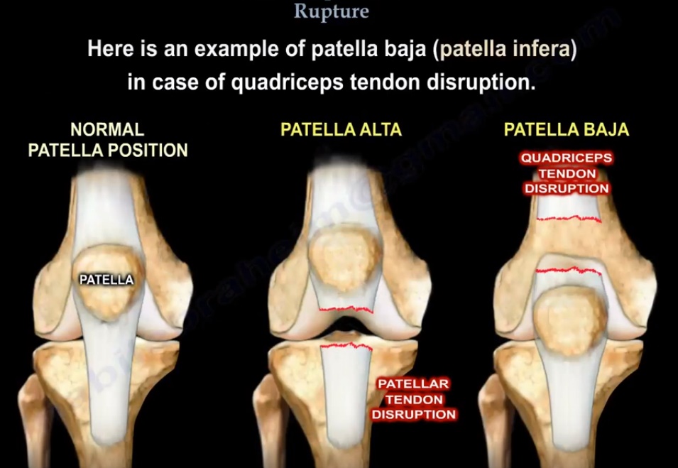
| Patellar position | Patella baja/infera | Patella alta |
| Mechanism | Failure of quadriceps tendon | Failure of patellar tendon |
| Extensor mechanism | Disrupted | Disrupted |
Important exam point
Quadriceps tendon rupture ? patella baja
Patellar tendon rupture ? patella alta
3. Predisposing Conditions
The lecture highlights several conditions associated with quadriceps tendon rupture:
- Rheumatoid arthritis
- Diabetes
- Hyperthyroidism
- Steroid injections around the knee
- Renal failure
- Gout
Bilateral quadriceps tendon rupture is uncommon but may occur, particularly in older patients.
Important point
Bilateral rupture should prompt evaluation for an underlying systemic disease.
4. Mechanism
The rupture occurs because of:
- Elongation and failure of the tendon
- Excessive loading of the knee extensor mechanism
The rupture may be:
- Partial
- Complete
5. Clinical Presentation
Patients may present with:
- Knee pain
- Swelling
- Extensor lag
- Difficulty walking
- Inability to actively extend the knee
- Inability to perform a straight-leg raise
Complete rupture
The quadriceps tendon is no longer attached effectively to the patella, resulting in loss of active knee extension.
Important clinical clue
Inability to perform a straight-leg raise strongly suggests disruption of the extensor mechanism.
6. Examination
A defect may be palpable:
- At the superior pole of the patella
- Usually within approximately 2 cm of the patella
If a defect is present but active extension is preserved
This suggests either:
- Partial quadriceps tendon tear, or
- Intact extensor retinaculum
The preserved retinaculum may allow some continuity of the extensor mechanism despite tendon disruption.
7. X-ray Findings
The characteristic radiographic finding is:
Patella baja / patella infera
The patella sits lower than its normal position.
Other findings may include:
- Loss of the normal quadriceps tendon shadow/mass
- Inferior position of the patella
Comparison
Quadriceps tendon rupture ? patella moves distally ? patella baja
Patellar tendon rupture ? patella moves proximally ? patella alta
8. MRI
MRI can clearly demonstrate the tendon injury.
It is particularly useful when:
- The diagnosis is uncertain
- It is unclear whether the tear is partial or complete
- The clinical examination is inconclusive
Important point
If the clinical picture is obvious, MRI may not be necessary before treatment.
9. Treatment
Complete Rupture
The lecture emphasizes early repair.
The reason for early treatment is to facilitate repair before the tendon becomes retracted or difficult to restore.
The tendon is repaired back to the superior pole of the patella.
10. Repair Technique
The described repair uses:
Non-absorbable sutures
- Running locking non-absorbable sutures
- Usually two sutures
- Produces four free suture ends
These sutures are passed through:
- Bone tunnels in the patella
The sutures are then tied to secure the quadriceps tendon to the patella.
Alternative fixation
Suture anchors may be used instead of transosseous bone tunnels.
11. Postoperative Immobilization
After repair:
- Splint or brace
- Approximately 4–6 weeks
The purpose is to protect the repaired extensor mechanism while healing occurs.
12. Complications
The lecture notes that after quadriceps tendon rupture and repair, the patient may experience approximately:
One-third loss of quadriceps muscle strength
Persistent weakness may therefore remain even after successful repair.
13. Bilateral Quadriceps Tendon Rupture
Bilateral rupture is rare.
When it occurs:
Look for an underlying systemic disease.
Particularly consider the metabolic and systemic conditions associated with tendon failure.
High-Yield Exam Points
- Quadriceps tendon rupture usually occurs in older patients, typically >40 years.
- More common in males.
- Usually occurs near the superior pole of the patella.
- Causes disruption of the extensor mechanism.
- Inability to straight-leg raise is an important clinical finding.
- Palpable defect is usually present above the patella.
- Quadriceps tendon rupture –patella baja/infera.
- Patellar tendon rupture – patella alta.
- MRI is particularly useful when distinguishing partial from complete rupture.
- Complete rupture should be repaired early.
- Repair can be performed using transosseous patellar tunnels or suture anchors.
- Postoperative immobilization is approximately 4–6 weeks.
- Bilateral rupture should prompt investigation for underlying systemic disease.
Leave a Reply