
Courtesy: Shan Nanji MD, www.kissanatomy.com Brachial Plexus Overview Brachial plexus arises from nerve roots C5–T1. Mnemonic for structure: ‘Reach To Drink Cold Beer’. R – Roots. T – Trunks. D – Divisions. C – Cords. B – Branches. Roots and Long Thoracic Nerve Roots: C5, C6, C7, C8, T1. Long thoracic nerve arises from C5–C7. […]
-Applied Anatomy
The Peroneus Tertius Muscle
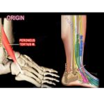
Courtesy: Prof Nabil Ebraheim, University of Toledo, Ohio, USA Peroneus tertius muscle The peroneus tertius muscle is absent in about 10% of the population. Located within the anterior compartment of the leg. Origin : from the distal third of the extensor surface of the fibula Insertion : into the base of 5th metatarsal bone Innervated […]
Anatomy Of The Sternoclavicular Joint
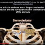
Courtesy: Prof Nabil Ebraheim, University of Toledo, Ohio, USA ANATOMY OF STERNOCLAVICULAR JOINT Composed Of proximal end of clavicle and manubrium sterni Clavicle in turn is supported by the costal cartilage of first rib Type : Gliding type/ saddle joint/ synovial joint Articular surface : proximal end of clavicle + clavicular notch of manubrium sterni […]
Anatomy of the Coracobrachialis and the Musculocutaneous Nerve
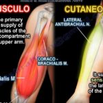
Courtesy: Prof Nabil Ebraheim, University of Toledo, Ohio, USA The coracobrachialis muscle arises from the tip of the coracoid process and inserts into the middle third of the medial border of humeral shaft. The muscle may have a conjoint tendon with the short head of the biceps muscle. The muscle lies lateral to the pectoralis […]
Sciatic Nerve Origin and Variations
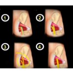
Courtesy: Prof Nabil Ebraheim, University of Toledo, Ohio, USA Sciatic nerve- origin, variations and course Origin Sciatic nerve is the largest nerve in the body. The Sciatic nerve arises from lumbosacral plexus in the pelvis. Sciatic nerve plus S4 is lumbosacral plexus The ventral rami of L4 to S3 nerve roots unite to form […]
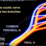
3D Anatomy of Sciatic Nerve
Courtesy: Prof Nabil Ebraheim, University of Toledo, Ohio, USA Overview Sciatic nerve is the largest nerve in the body. Originates from the lumbosacral plexus (L4–S3 nerve roots). Formed by two components: tibial nerve and common peroneal (fibular) nerve. Course of the Sciatic Nerve Leaves the pelvis through the greater sciatic foramen. Usually passes inferior to […]
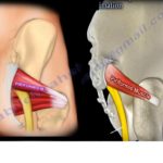
Anatomy of Distal Brachial Plexus

Courtesy: Prof Nabil Ebraheim, University of Toledo, Ohio, USA Overview The brachial plexus is the nerve network that supplies motor and sensory innervation to the upper limb. It is formed by the anterior rami of five spinal nerves. These spinal nerve roots combine and reorganize to form trunks, divisions, cords, and finally the terminal nerves […]
Anatomy of the Triceps Muscle
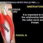
Courtesy: Prof Nabil Ebraheim, University of Toledo, Ohio, USA Triceps muscle is a powerful extensor of the elbow joint -has 3 heads . Long head . Lateral head . Medial head Long and lateral head forms superficial part of triceps Medial head forms deep part of triceps Long head Arise from the scapula and stretches […]
Q angle of the Knee Joint

Courtesy: Prof Nabil Ebraheim, University of TOledo, Ohio, USA A well functioning knee joint is important for mobility and to support the weight of the body during day to day activities .Hence normal alignment of the knee is essential for its proper functioning. Q angle (Quadriceps angle )is the angle between the quadriceps tendon and […]
Anatomy of the Pronator Teres Muscle
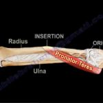
Courtesy: Prof Nabil Ebraheim, University of Toledo, Ohio, USA ANATOMY OF PRONATOR TERES MUSCLE • Muscle located in the forearm • Origin: from 2 heads- Superficial and deep head Superficial head which is the humeral head arises from medial epicondyle of the humerus. Deep head which is the ulnar head arises from the medial […]
Anatomy of Serratus Anterior Muscle
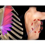
Courtesy: Prof Nabil Ebraheim, University of Toledo, Ohio, USA The serratus anterior is a fan-shaped muscle that originates on the superolateral surfaces of the first to eighth ribs and inserts along the anterior aspect of medial border of the scapula. It is the strong protractor of scapula . It is known as BOXERS MUSCLE […]
The Lumbar Plexus Simplified
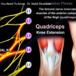
Courtesy: Prof Nabil Ebraheim, University of Toledo, Ohio, USA Overview The lumbosacral plexus is a network of nerves supplying the lower limb and pelvic region. The lumbar plexus forms the upper portion of this network. It arises primarily from the anterior rami of spinal nerves L1 to L4, with occasional contribution from T12. Part of […]
Anatomy of the Lumbosacral Plexus
Courtesy: Prof Nabil Ebraheim, University of Toledo, Ohio, USA Overview The lumbosacral plexus is a network of nerves supplying the lower limb, gluteal region, and perineum. It is formed by the combination of lumbar and sacral nerve roots. The sciatic nerve is the most important and largest nerve within this plexus. Understanding the sciatic nerve […]
Anatomy of Profunda Femoris Artery
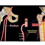
Courtesy: Prof Nabil Ebraheim, University of Toledo, Ohio, USA The Profunda femoris artery which is the chief blood supply of the thigh,arises from the posterolateral aspect of the femoral artery about 4cm below the inguinal ligament.Speaking about its course,it passes medially behind the femoral artery.It crosses the Pectineus and the Adductor brevis muscles,runs under the […]
Anatomy of Lateral Femoral Circumflex Artery
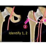
Courtesy: Prof Nabil Ebraheim, University of Toledo, Ohio, USA Lateral Circumflex Femoral Artery The lateral circumflex femoral artery is a branch of the profunda femoris artery. The profunda femoris artery is the main blood supply to the thigh. The profunda femoris artery gives two circumflex arteries and four perforating branches. The two circumflex arteries […]