Courtesy: Prof Nabil Ebraheim, University of TOledo, Ohio, USA
A well functioning knee joint is important for mobility and to support the weight of the body during day to day activities .Hence normal alignment of the knee is essential for its proper functioning.
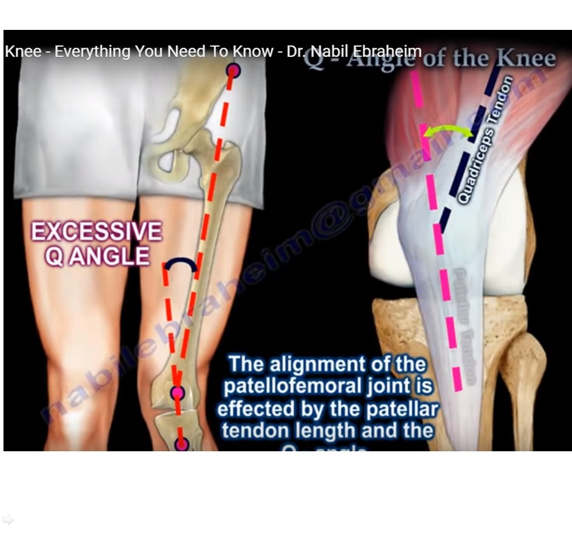
Q angle (Quadriceps angle )is the angle between the quadriceps tendon and the patellar tendon . This angle provides useful information regarding the alignment of the knee joint.Any increase in the angle is considered to be a risk factor for patellar subluxation .
Q angle is formed in the frontal plane by two line segments . First line is drawn from the anterior superior iliac spine to the centre of the patella.Second line is drawn from the centre of the patella to the tibial tubercle.The angle formed by the two lines is known as the Q angle.Its always best to measure the angle with the knee in extension as well as flexion . Normal Q angle is variable .In extension ,the normal Q angle for males is usually 14 ° and for females is usually 17°.In flexion ,the normal Q angle is approximately 8°.
The alignment of the patellofemoral joint is effected by the patellar tendon length and the Q angle.A wider pelvis in females and an increase in Q angle is linked to knee pain ,patellofemoral pain and ACL injury . Increased Q angle is mostly seen in genu valgum, external tibial torsion ,femoral anteversion,lateral positioned tibial tuberosity ,tight lateral retinaculum. Large Q angle and a strong quadriceps contraction can dislocate the patella. CT scan study of the patellofemoral articulation is helpful .
An abnormal patellar tracking is one of the most common complication of total knee replacement.Preservation of a normal Q angle is important .An increase in the Q angle in the knee will lead to an increase in the lateral subluxation forces on the patella , which may lead to pain,tear of the implant ,and mechanical symptoms .When doing a total knee replacement , avoid techniques that will cause an increase in Q angle such as internal rotation of the femoral component or the tibial component or avoid medialization of the femoral component , avoid lateral placement of the patellar component.The patellar prosthesis should be placed either in the centre or slightly medial .
Miserable malignant syndrome – It is a term used to describe a triad of anatomic features or finding which will cause an increase in the Q angle.It includes excessive femoral anteversion,genu valgum , external tibial torsion or pronated feet .The symptoms include anterior knee pain ,pain under the patella , stiffness of the knee joint . When examining the patient for patellofemoral pain ,the alignment is important including the rotational alignment .

Leave a Reply