Courtesy: Prof Nabil Ebraheim, University of Toledo, Ohio, USA
The lumbosacral plexus is a network of nerves supplying the:
- Lower limb
- Pelvic region
The lumbar plexus forms the upper part of this system.
Key Features
- Arises from anterior rami of L1–L4
- May receive contribution from T12
- Part of L4 forms the lumbosacral trunk, connecting to the sacral plexus
Lumbar Plexus
Origin
Formed from:
- L1
- L2
- L3
- L4
- ± Contribution from T12
Branches of the Lumbar Plexus
Six Main Nerves
- Iliohypogastric nerve
- Ilioinguinal nerve
- Genitofemoral nerve
- Lateral femoral cutaneous nerve
- Obturator nerve
- Femoral nerve
Memory Aid
“I Twice Got Lunch On Friday”
| Letter | Nerve |
|---|---|
| I | Iliohypogastric |
| I | Ilioinguinal |
| G | Genitofemoral |
| L | Lateral femoral cutaneous |
| O | Obturator |
| F | Femoral |
Nerve Root Contributions
From One Root
- Iliohypogastric – L1
- Ilioinguinal – L1
From Two Roots
- Genitofemoral – L1–L2
- Lateral femoral cutaneous – L2–L3
From Three Roots
- Obturator – L2–L3–L4
- Femoral – L2–L3–L4
Relation to Psoas Major Muscle
Lateral Border (Most Nerves)
- Iliohypogastric
- Ilioinguinal
- Lateral femoral cutaneous
- Femoral
Anterior Surface
- Genitofemoral nerve
Medial Border
- Obturator nerve
Major Lumbar Plexus Nerves
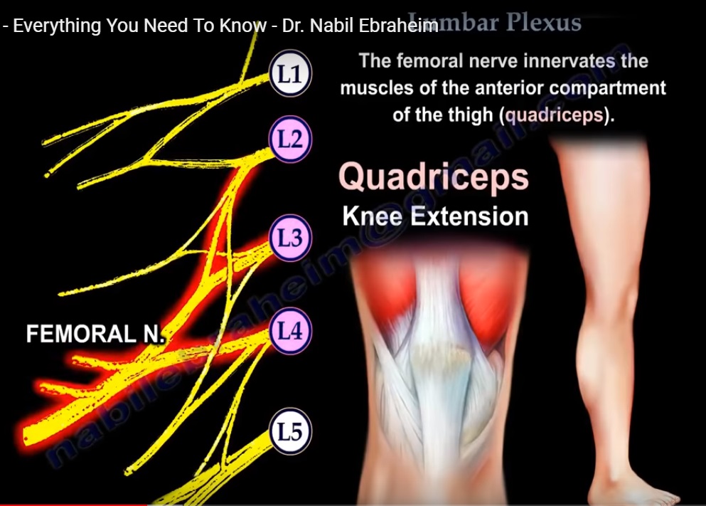
Femoral Nerve
Roots
- L2–L4
Motor Function
- Innervates quadriceps
- Responsible for knee extension
Sensory Supply
- Anterior thigh
- Medial leg (via saphenous nerve)
Clinical Relevance
- Weak knee extension
- Reduced patellar reflex
Obturator Nerve
Roots
- L2–L4
Motor Function
- Supplies adductor muscles
- Responsible for hip adduction
Sensory Supply
- Medial thigh
Lateral Femoral Cutaneous Nerve
Roots
- L2–L3
Type
- Purely sensory
Function
- Sensation over lateral thigh
Clinical Relevance
- Compression – Meralgia paresthetica
Genitofemoral Nerve
Roots
- L1–L2
Branches
- Genital branch
- Femoral branch
Functions
- Genital branch:
- Cremaster muscle
- Genital skin
- Femoral branch:
- Upper anterior thigh sensation
Iliohypogastric Nerve
Root
- L1
Function
- Supplies:
- Abdominal wall muscles
- Lower abdomen
- Upper lateral gluteal region
Ilioinguinal Nerve
Root
- L1
Function
- Supplies:
- Abdominal wall
- Upper medial thigh
- External genitalia
Key Summary Points
Structural Overview
- Lumbar plexus arises from L1–L4 (± T12)
- Forms the upper component of the lumbosacral plexus
Branch Pattern
- 2 nerves from one root
- 2 nerves from two roots
- 2 nerves from three roots
Important Clinical Nerves
- Femoral nerve — anterior thigh
- Obturator nerve — medial thigh
Relation to Psoas Major
- Most nerves emerge laterally
- Exceptions:
- Genitofemoral — anterior
- Obturator — medial
Clinical Insight
Understanding this organized pattern helps in:
- Neurological localization
- Diagnosis of nerve injuries
- Surgical planning
Leave a Reply