
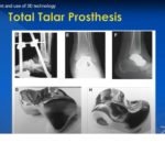
Courtesy: Chayannin Angthong MD, Thammasat University, Thailand
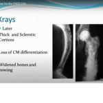
Bone Tumours for the FRCSOrth

Courtesy: Mostafa Elgendy, Jeddah, Saudi Arabia
Metabolic Bone disease for the FRCSOrth
Courtesy: Dr Rohan Bidwai, FRCS Tr and Orth, UK
Anatomy Of The Latissimus Dorsi Muscle
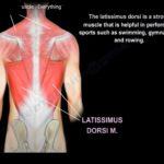
Courtesy: Prof Nabil Ebraheim, University of Toledo, Ohio, USA Anatomy of the Latissimus Dorsi Muscle Latissimus dorsi muscle is anatomically is a muscle on the back but functionally is a muscle on the upper limb. It is the broadest muscle of the back. It is one of the strongest muscle and is used in sports […]
Anatomy of the Trapezius Muscle
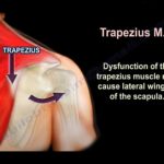
Courtesy: Prof Nabil Ebraheim, University of Toledo, Ohio, USA Anatomy of Trapezius muscle Trapezius is a large superficial muscle that extends from the back of the skull,back of the neck and back of the thorax. Origin: The upper fibres of the trapezius muscle arise from the external occipital protruberance and the medial third of the […]
Concise Orthopaedic Notes: Revision aid for FRCS ,EBOT Board Examinations
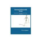
ISBN: 1696747872 Ebook and paperback:751 pages Editor:Firas Arnaout The authors launch the 2nd edition of their highly popular book ‘Concise Orthopaedic Notes’ first published in 2019. The original edition has already been awarded the winner in the ‘Best new orthopaedic book’ category by BookAuthority. This book is extremely useful for any professional involved in trauma […]
Differential Diagnosis of Lateral Elbow Pain

Courtesy: Dr Michelle Ruiz Suares, President Mexico Shoulder and Elbow Society, Mexico city, Mexico Elbow Pain – Causes, Diagnosis, and Treatment Overview Elbow pain is a common clinical problem Causes vary widely — accurate diagnosis is essential Key Concept Pain is best classified based on: Location Classification Based on Pain Location 1. Anterior (Front) […]
Scoliosis for the FRCSOrth exam
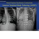
Courtesy: Muralidharan Venkatesan, FRCS Tr and Orth, Apollo Hospitals, Chennai
Paediatric Upper extremity Trauma
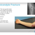
Courtesy: Amr Abdelgawad, Maimonaides Medical Centre, NY, USA Pediatric Upper Extremity Trauma 1. Medial Epicondyle Fracture (Children) Key Anatomy Last ossification center to fuse in elbow Fusion age: Boys: 16–19 years Girls: 13–16 years Attachments Medial collateral ligament (MCL) Mechanism Valgus stress – avulsion injury Commonly associated with elbow dislocation Treatment Non-operative (Most Cases) […]
Patellofemoral Instability

Courtesy: Yogesh Joshi, Consultant Knee Surgeon, Wrexham, UK
Spine- Pelvis Relationship in THR
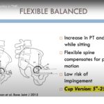
Courtesy: Savyasachi Thakkar, Asst Professor, John Hopkins Medical Centre, Baltimore, USA
Ankle Fractures – An Evidence Based Review for FRCS

Courtesy: Prof Lyndon Mason, Liverpool, UK
Asceptic Loosening and Wear characteristics for the FRCS Orth

Courtesy: Ajith Appuhamy, FRCS Tr and Orth
Anatomy Of The Teres Minor Muscle
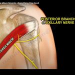
Courtesy: Prof Nabil Ebraheim, University of Toledo, Ohio, USA Anatomy of the teres minor muscle The teres minor is a narrow muscle which originates from the posterior lateral surface of the scapula and inserts into the greater tuberosity of the humerus . Teres minor is innervated by the posterior branch of axillary nerve . The […]
Realignment Osteotomies for Arthritic Knee
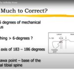
Courtesy: KrishnaKumar, Limb Reconstruction Surgeon, Trichur, Kerala, India Correction of Extra-articular Deformity in Total Knee Arthroplasty 1. Basic Concept Significant extra-articular deformity around the knee is relatively uncommon in some practices, but may be encountered following: Malunited fractures Previous osteotomies with residual malalignment Metabolic bone disease When performing TKA in the presence of extra-articular […]