
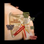
Courtesy: Prof Nabil Ebraheim, University of Toledo, Ohio, USA Anatomy of Teres Major Muscle Origin- dorsal aspect of inferior angle of scapula Insertion- medial lip of intertubercular groove of the humerus One of the muscle that connect scapula to the humerus Teres major muscle does not attach to the capsule of glenohumeral joint Teres major […]
Radial Nerve Injuries for the FRCSOrth Exam
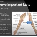
Courtesy: Santhosh Venkatachalam, Northumbria Healthcare, UK
Adult FlatFoot Deformity
Courtesy: Amr Abdelgawad, Maimonaides Medical Centre, NY, USA
Overview of Paediatric Upper Extremity Trauma
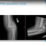
Courtesy: Amr Abdelgawad, Maimonaides Medical Centre, NY, USA Pediatric Upper Extremity Trauma Clavicle Fracture Key anatomy First bone to ossify during fetal development. Medial clavicular epiphysis is the last epiphysis to fuse at 20 to 25 years. Management Children younger than 12 years are treated nonoperatively in almost all cases. Adolescents 15 years and […]
Overview of Paediatric Lower Extremity Trauma
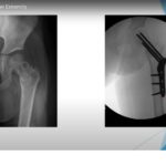
Courtesy: Amr Abdelgawad, Maimonaides Medical Centre, NY, USA Apophyseal and Avulsion Injuries Apophysis Secondary ossification center located outside the joint where tendons attach. Common apophyseal attachments Calcaneus: Achilles tendon. Tibial tubercle: Patellar tendon. Anterior inferior iliac spine (AIIS): Rectus femoris. Anterior superior iliac spine (ASIS): Sartorius. Ischial tuberosity: Hamstrings. Iliac crest: Abdominal muscles. Treatment Rest. […]
Research Basics
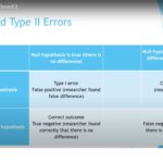
Courtesy: Amr Abdelgawad, Maimonaides Medical Centre, NY, USA
Slipped Capital Femoral Epiphysis
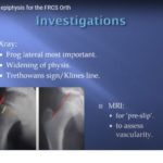
Courtesy: Sally Hobson, Hull Royal Infirmary, UK Definition Slipped capital femoral epiphysis is a Salter–Harris Type I injury occurring through the hypertrophic zone of the proximal femoral growth plate. The femoral head remains within the acetabulum, while the femoral neck and shaft displace superolaterally and externally rotate relative to the epiphysis. Epidemiology Commonly occurs between […]
Achilles Tendinopathy for the FRCS Orth

Courtesy: Anjani Kumar Singh, Liverpool, UK
Scaphoid fracture, SNAC and SLAC wrist
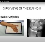
Courtesy: Saurabh Agarwal, London, UK
Peroneal Tendon Tears and Dislocation

Courtesy: Kurt Hofmann MD, New England Baptist Hospital, Boston, Massachussets, USA
Anatomy and Blood supply of Talus
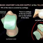
Courtesy: Prof Nabil Ebraheim, University of Toledo, Ohio, USA Basic Anatomy of the Talus Approximately 70% of the talus surface is covered by articular cartilage. Talus consists of head, neck, body, lateral process, and posterior process. Large cartilage coverage contributes to high risk of post?traumatic arthritis after fractures. Blood Supply of the Talus Primary blood […]
Ochronosis and Alkaptonuria
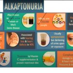
Courtesy: Dr Shivshankar Challa, MS, MRCS, Mch Introduction: Overview of Alkaptonuria Alkaptonuria is an extremely rare autosomal recessive metabolic disorder. It is caused by deficiency of the enzyme homogentisic acid dioxygenase. The condition affects approximately one in two hundred and fifty thousand to one in one million live births. Under normal metabolism, homogentisic acid is […]
Matrix Encapsulated Autologous Chondrocyte Implantation
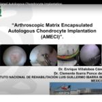
Courtesy: By Dr Enrique Villalobos Cordova, Instituto Nacional de Rehabilitación Luis Guillermo Ibarra Ibarra, Mexico city, Mexico
Kanavel’s Signs

Courtesy: Prof Nabil Ebraheim, University of Toledo, Ohio, USA
Bone and Cartilage Histology, Collagen
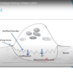
Courtesy: Amr Abdelgawad, Maimonaides Medical Centre, NY, USA Types of Bone Organized (Lamellar) Bone Mature, well-organized bone Provides strength and structural stability Cortical Bone Location Forms the outer cortex of long bones Predominantly seen in the diaphysis (shaft) Characteristics Dense with low porosity Low surface area High strength and stiffness Lower metabolic activity Cancellous (Trabecular) […]