
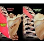
Courtesy: Prof Nabil Ebraheim, University of Toledo, Ohio, USA ANATOMY, FUNCTION AND DYSFUNCTION OF RHOMBOID MUSCLES Rhomboid muscles are rhombus shaped muscles that lie underneath the trapezius muscle They connect scapula to the vertebrae and thus holds it close to the thoracic wall It is shaped like a diamond or kite, i.e a rhombus […]
Anatomy of the Adductor Longus Muscle
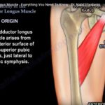
? Courtesy: Prof Nabil Ebraheim, University of Toledo, Ohio, USA ADDUCTOR LONGUS One of the six adductor muscles located within the thigh, the others being pectineus, adductor brevis, adductor magnus, gracilis and obturator externus Origin Arises from the anterior surface of the superior pubic ramus, lateral to the pubic symphysis Insertion Middle third of medial […]
Posterior Malleolar Fractures- Why Should we Care?
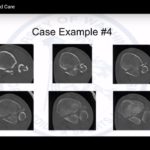
Courtesy: Sean E. Nork, MD, Professor, Harborview Medical Center, Department of Orthopaedics and Sports Medicine University of Washington School of Medicine Claudia Christman-Skieller, MD (R4) Orthopaedic Surgery Resident Department of Orthopaedics and Sports Medicine University of Washington School of Medicine
Ulnar Styloid fractures
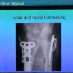
Courtesy: Vikas Gupta, Max Hospital, Saket, Gurgaon, India
Anatomy Of The Pectineus Muscle
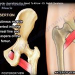
Courtesy: Prof Nabil Ebraheim, University of Toledo, Ohio, USA ANATOMY OF PECTINEUS MUSCLE Pectineus muscle is a flat quadrangular muscle situated at the anterior part of upper medial aspect of thigh It is one of the six adductors of thigh ORIGIN From superior pubic ramus INSERTION Into pectineal line and linea aspera of femur NERVE […]
Volar and Dorsal Barton fractures
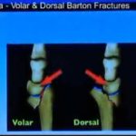
Courtesy: Dr Vikas Gupta, Consultant Hand Surgeon, Max Hospital, Saket, Gurgaon, India
Anatomy Of The Semimembranosus Muscle
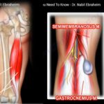
Courtesy: Prof Nabil Ebraheim, University of Toledo, Ohio, USA Anatomy of Semimembranosus One of the 3 muscles that make up the hamstring. The other 2 are , semitendinosus and biceps femoris Origin Upper lateral part of ischial tuberosity Insertion Back of the posterior surface of the medial tibial condyle Nerve supply Sciatic nerve Action 1.Flexion […]
Anatomy of the Semitendinosus Muscle
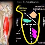
Courtesy: Prof Nabil Ebraheim, University of Toledo, Ohio, USA Semitendinosus is one of the three muscles that make up the hamstrings muscle group, and it is located at the posterior and medial aspect of the thigh The hamstring muscles are- semitendinosus, semimembranosus and biceps femoris. It is a cylinder muscle- starts and ends as tendon […]
Evolution of fixation of distal radius fractures
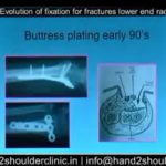
Courtesy: Dr Vikas Gupta, Max Hospital, Saket, Gurgaon, India
The Ponseti Method Of Clubfoot Care

Courtesy: Dr. John Norgrove Penny and www.global-help.org
Treatment of Mallet Finger
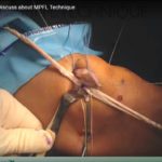
Courtesy: Mathew Ricks, Ashok, Shyam, IORG, OrthoTV
Understanding and Analyzing Gait For The Clinician
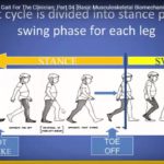
Courtesy:Michael Sussman MD and www.global-help.org
Computer Based 3D analysis of Gait
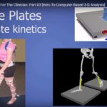
Courtesy: MIchael Sussman MD, and www.global-help.org Three-Dimensional Gait Analysis Overview 3D gait analysis is an advanced method to study human walking objectively Widely used in: Research Outcome studies Surgical planning Clinical Relevance Helps evaluate: Treatment outcomes Functional improvement Disease progression Key Concept Even without access to a gait lab: Clinical observation remains essential Definition […]
Shattered Lower end Radius fractures
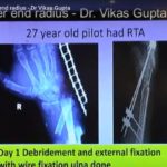
Courtesy: Dr Vikas Gupta, Max Hospital, Saket, Gurgaon, India
Rectus sparing Periacetabular Osteotomy
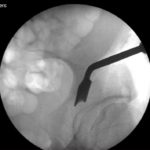
Courtesy: Christopher Peters MD, University of Utah, USA