Courtesy: MIchael Sussman MD, and www.global-help.org
Three-Dimensional Gait Analysis
Overview
- 3D gait analysis is an advanced method to study human walking objectively
- Widely used in:
- Research
- Outcome studies
- Surgical planning
Clinical Relevance
- Helps evaluate:
- Treatment outcomes
- Functional improvement
- Disease progression
Key Concept
- Even without access to a gait lab:
- Clinical observation remains essential
Definition of Gait Analysis
- Gait analysis = study of walking by breaking it into measurable components
Three Anatomical Planes
1. Sagittal Plane
- Forward–backward movement
- Examples:
- Hip flexion/extension
- Knee flexion/extension
2. Coronal Plane
- Side-to-side movement
- Examples:
- Hip abduction/adduction
3. Transverse Plane
- Rotational movement
- Examples:
- Pelvic rotation
- Limb rotation
Purpose of 3D Gait Analysis
- Assist in surgical decision-making
- Evaluate postoperative outcomes
- Monitor progression over time
- Objectively assess improvement or deterioration
Evolution of Gait Analysis
Earlier Systems (1970s)
- Time-consuming
- Took days for analysis
Modern Systems
- Complete test: ~2 hours
- Data processing: 30–45 minutes
- Digital storage for review
Components of a Full Gait Lab Evaluation
1. Physical Examination
- Performed before gait testing
- Includes:
- Range of motion
- Muscle strength
- Limb alignment
- Functional status
2. Video Analysis
- High-quality recording of walking
- Helps visualize gait abnormalities
- Duration: 15–20 minutes
3. Marker Placement
- Reflective markers placed on:
- Limbs
- Pelvis
- Trunk
Purpose
- Identify joint centers
- Track movement
4. Motion Capture System
- Patient walks in room with 6–12 infrared cameras
Function
- Detect marker movement
- Calculate 3D position
Accuracy
- Requires >/= 2 cameras per marker
Output
- Stick-figure reconstruction
- ~120 frames/second recording
Gait Graphs
Graph Layout
| Column | Plane | Movement |
|---|---|---|
| 1 | Sagittal | Flexion/extension |
| 2 | Coronal | Side-to-side |
| 3 | Transverse | Rotation |
Reference Values
- Gray bands represent:
- Normal range (mean ± SD)
Linear Gait Parameters
Important Measures
- Cadence
- Steps per minute
- Step Length
- Distance between opposite heel strikes
- Stride Length
- Distance in one full gait cycle
- Velocity
- Walking speed (m/s)
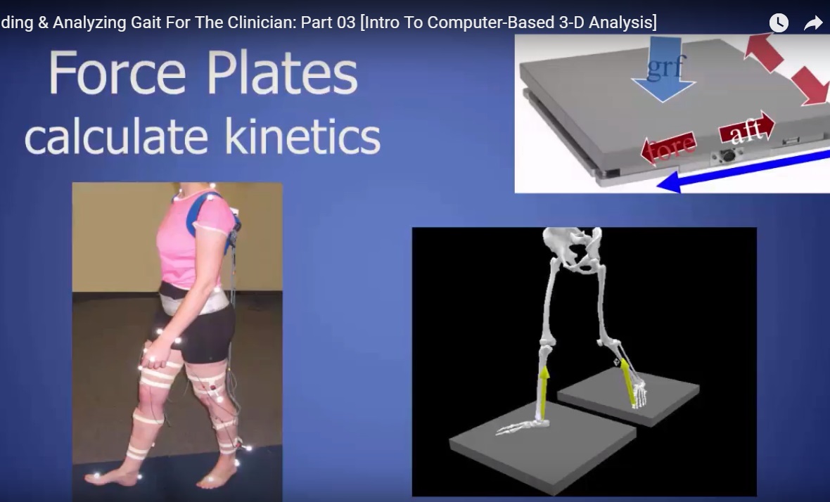
Kinetics (Force Analysis)
Measured Using Force Plates
Forces Assessed
- Vertical Force
- Similar to body weight
- Anterior–Posterior Force
- Forward/backward forces
- Medial–Lateral Force
- Side-to-side forces
Clinical Use
- Calculates:
- Joint forces
- Joint moments
Dynamic Electromyography (EMG)
Purpose
- Measures muscle activity during gait
Method
- Surface electrodes placed on muscles
- Typically 8 channels
Special Cases
- Needle EMG for deep muscles
Interpretation
- Shows:
- Timing of muscle activation
- Does NOT measure:
- Strength of contraction
Clinical Importance
- Useful in neurological conditions (e.g., cerebral palsy)
- Detects abnormal continuous muscle activity
Foot Pressure Analysis
1. Static Analysis
- Patient stands on platform
- Evaluates:
- Foot alignment
- Plantar surface
Limitation
- No dynamic information
2. Dynamic Analysis (Pedobarography)
Measures
- Pressure distribution during walking
Provides
- Center of pressure pathway
- Load progression:
- Heel — midfoot — forefoot — toes
Clinical Example: Clubfoot Follow-Up
Case
- 10-year-old child treated for clubfoot
Findings
- Supination during stance
- Abnormal pressure distribution
Treatment
- Tibialis anterior tendon transfer
Outcome
- Improved load distribution
- Better forefoot function
Key Points
- 3D gait analysis provides objective biomechanical data
- Combines:
- Motion analysis
- Force measurement
- EMG
- Pressure studies
Final Message
- Despite advanced technology:
- Clinical examination remains the cornerstone of gait assessment
Leave a Reply