Courtesy: Prof Young Lae Moon, Seoul, South Korea Comprehensive Shoulder Physical Examination Orthopaedic Principles – Webinar Summary 1. Introduction A systematic shoulder examination is essential for accurate diagnosis and treatment planning. A structured, stepwise approach improves diagnostic precision and reduces missed pathology. Key focus: Cervical spine assessment Rotator cuff evaluation Shoulder instability assessment 2. Core […]
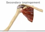
Tennis Elbow
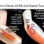
? Courtesy: Prof Nabil Ebraheim, University of Toledo, Ohio, USA Tennis Elbow (Lateral Epicondylitis) Definition Tennis elbow, also called lateral epicondylitis, is an overuse injury. It causes: Inflammation Tendinosis Lateral elbow pain The pathology occurs at the origin of the Extensor Carpi Radialis Brevis (ECRB) muscle. Pain is located on the outer (lateral) side of […]
Baker’s Cyst
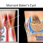
Courtesy: Prof Nabil Ebraheim, University of Toledo, Ohio, USA Baker’s Cyst (Popliteal Cyst) Definition Baker’s cyst, also called a popliteal cyst, is a benign swelling located behind the knee. It may vary in size, from small to large. It is a fluid-filled cyst, not a solid tumor. The cyst should transilluminate. Anatomy and Pathophysiology The […]
Common Nerve Conditions of the Upper Extremity
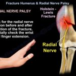
Courtesy: Prof Nabil Ebraheim, University of Toledo, Ohio, USA 1. Nerve Injuries in Supracondylar Fracture of the Humerus (Children) Supracondylar fractures of the humerus in children are commonly associated with nerve injuries. Extension Type (Most Common) Most common type of supracondylar fracture. Commonly associated with: Anterior interosseous nerve (AIN) palsy It is essentially a high […]
Nerve Injuries of the Lower Extremity
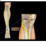
Courtesy: Prof Nabil Ebraheim, University of Toledo, Ohio, USA 1. Tarsal Tunnel Syndrome Overview One of the most common nerve injuries in the lower extremity. Approximately 80% of cases have no identifiable cause. Involves compression of the posterior tibial nerve. Symptoms Burning sensation Numbness Tingling Electric shock–like pain Symptoms occur on the plantar (bottom) aspect […]
Understanding Ulnar Nerve Facts
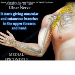
? Courtesy: Prof Nabil Ebraheim, University of Toledo, Ohio, USA 1. Causes of Ulnar Nerve Compression The most common cause: Compression between the two heads of the flexor carpi ulnaris. Other causes include: Anconeus epitrochlearis muscle. Arcade of Struthers. Differentiate between: Arcade of Struthers – may compress the ulnar nerve. Ligament of Struthers – associated […]
Rotator Cuff Pathology
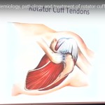
Courtesy: Prof Nabil Ebraheim, University of Toledo, Ohio, USA 1. Anatomy of the Rotator Cuff The rotator cuff consists of four muscles: Supraspinatus Infraspinatus Teres minor Subscapularis Each muscle has a specific function in shoulder movement and stability. 2. Rotator Cuff Tears Common source of shoulder pain. May involve: A single tendon Multiple tendons Tears […]
Antalgic Gait
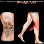
Courtesy: Prof Nabil Ebraheim, University of Toledo, Ohio, USA Definition Antalgic gait is a painful gait pattern. The patient avoids spending time on the affected leg due to pain. The goal is to reduce weight-bearing on the painful extremity. Normal Gait Cycle Stance phase: 60% of the gait cycle Swing phase: 40% of the gait […]
Achilles Tendon Pathology
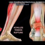
Courtesy: Prof Nabil Ebraheim, University of Toledo, Ohio, USA 1. Location and Structure The Achilles tendon is located in the posterior ankle. It is the strongest and thickest tendon in the body. Formed from: Soleus muscle Gastrocnemius muscle Inserts into the calcaneus (heel bone). 2. Associated Bursae Subcutaneous Calcaneal Bursa Located superficial to the Achilles […]
Low Back Pain
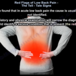
Courtesy: Prof Nabil Ebraheim, University of Toledo, Ohio, USA Importance of Identifying Red Flags A detailed history and careful physical examination are essential. Red flags are warning signs that may indicate a serious underlying condition. In the absence of red flags, acute low back pain is usually treated conservatively. Management of Uncomplicated Acute Low Back […]
Jones Fracture
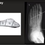
Courtesy: Prof Nabil Ebraheim, University of Toledo, Ohio, USA Metatarsal Fractures Metatarsal fractures can involve: First metatarsal Fifth metatarsal Second, third, and fourth metatarsals (including stress fractures) 1. First Metatarsal Fractures Key Points Different from fractures of the second, third, and fourth metatarsals. The first metatarsal carries a greater load. Malunion may cause: Transfer lesions […]
Shoulder Arthroplasty: Current Concepts

Courtesy: Prof William Levine, Past President, ASES Historical Evolution of Shoulder Arthroplasty Professor Levine outlined six innovation eras. Era 1: Early Shoulder Arthroplasty (Late 1800s–1950s) Themistocles Gluck (Germany) First shoulder arthroplasty Modular ivory prosthesis Not widely recognized at the time Jules Péan (France) First documented shoulder replacement (1893) Two-stage procedure Ultimately failed due to infection […]
Cartilage Injuries of the Knee
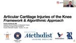
Courtesy: Sachin Allahabadi MD, Houston, TX Why Do Cartilage Defects Matter? 1. Poor Healing Potential Articular cartilage has: Limited blood supply Low chondrocyte density Dense extracellular matrix Limited cell migration capacity Additionally: The knee undergoes repetitive multidirectional load Healing is mechanically and biologically challenging 2. High Prevalence Articular cartilage defects are found in ~60% of […]
Bones that trick you: Fracture Mimics

Courtesy: Prof Shital Parikh MD, Cincinnati Childeren’s Hospital, Philadelphia, PN Part 1: Fracture Mimickers in Children 1. Irregular Ossification – A Common Trap Pediatric ossification centers are irregular and may mimic fractures. The elbow is particularly challenging due to multiple ossification centers. Familiarity with: Order of appearance Timing of fusion Normal variantsis essential to avoid […]
Medial Collateral Ligament Injuries
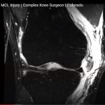
Courtesy: Prof Wolf Petersen, Martin Luther Krakenhaus, Berlin, Germany Anatomy of the Medial Collateral Ligament Complex The medial side of the knee consists of three principal ligamentous structures: 1. Superficial Medial Collateral Ligament Origin: Medial femoral epicondyle Insertion: Approximately 7–8 centimeters distal to the joint line, below the pes anserinus Function: Primary restraint to valgus […]