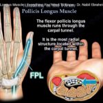
Courtesy: Prof Nabil Ebraheim, University of Toledo, Ohio, USA Video describes the Anatomy of Flexor Pollicis Longus Muscle. It is one of the three deep flexors muscles of the forearm. The Flexor Pollicis Longus Muscle Originates from the anterior surface of the Radius and the Interrosseus membrane. It runs through the carpal tunnel and get […]
-Applied Anatomy
Anatomy Of The Gastrocnemius Muscle
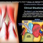
Courtesy: Prof Nabil Ebraheim, University of Toledo, Ohio, USA Video describes The Anatomy of Gastrocnemius Muscle. It arises from femur and crosses the knee joint and Ankle joint.It is part of the three Superficial Flexors in the leg. The Gastrocnemius Muscle has 2 heads, one medial and one lateral head. The Medial head originates from […]
Anatomy Of The Adductor Brevis Muscle
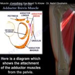
Courtesy: Prof Nabil Ebraheim, University of Toledo, Ohio, USA This video describes the Anatomy of the adductor Brevis Muscle which is one of the Six adductors of The Hip. The Muscle Originates from the inferior Pubic Ramus, inferior to the origin of Adductor Longus Muscle.It is Inserted into the Pectineal line and Superior part of […]
Anatomy, Function and Dysfunction of Rhomboid Muscles
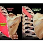
Courtesy: Prof Nabil Ebraheim, University of Toledo, Ohio, USA ANATOMY, FUNCTION AND DYSFUNCTION OF RHOMBOID MUSCLES Rhomboid muscles are rhombus shaped muscles that lie underneath the trapezius muscle They connect scapula to the vertebrae and thus holds it close to the thoracic wall It is shaped like a diamond or kite, i.e a rhombus […]
Anatomy of the Adductor Longus Muscle
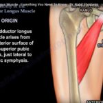
? Courtesy: Prof Nabil Ebraheim, University of Toledo, Ohio, USA ADDUCTOR LONGUS One of the six adductor muscles located within the thigh, the others being pectineus, adductor brevis, adductor magnus, gracilis and obturator externus Origin Arises from the anterior surface of the superior pubic ramus, lateral to the pubic symphysis Insertion Middle third of medial […]
Anatomy Of The Pectineus Muscle
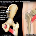
Courtesy: Prof Nabil Ebraheim, University of Toledo, Ohio, USA ANATOMY OF PECTINEUS MUSCLE Pectineus muscle is a flat quadrangular muscle situated at the anterior part of upper medial aspect of thigh It is one of the six adductors of thigh ORIGIN From superior pubic ramus INSERTION Into pectineal line and linea aspera of femur NERVE […]
Anatomy Of The Semimembranosus Muscle
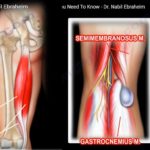
Courtesy: Prof Nabil Ebraheim, University of Toledo, Ohio, USA Anatomy of Semimembranosus One of the 3 muscles that make up the hamstring. The other 2 are , semitendinosus and biceps femoris Origin Upper lateral part of ischial tuberosity Insertion Back of the posterior surface of the medial tibial condyle Nerve supply Sciatic nerve Action 1.Flexion […]
Anatomy of the Semitendinosus Muscle
Courtesy: Prof Nabil Ebraheim, University of Toledo, Ohio, USA Semitendinosus is one of the three muscles that make up the hamstrings muscle group, and it is located at the posterior and medial aspect of the thigh The hamstring muscles are- semitendinosus, semimembranosus and biceps femoris. It is a cylinder muscle- starts and ends as tendon […]
Anatomy Of The Acetabulum
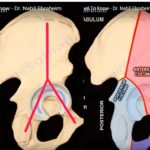
Courtesy: Prof Nabil Ebraheim, University of Toledo, Ohio, USA Overview The acetabulum is an important component of the hip joint and forms the socket for the femoral head. For clinical and surgical purposes, the acetabulum is divided into two structural columns. Understanding these columns is essential for interpreting acetabular fractures and planning surgical treatment. Several […]
Anatomy of Flexor Digitorum Superficialis
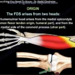
Courtesy: Prof Nabil Ebraheim, University of Toledo, Ohio, USA ANATOMY OF FLEXOR DIGITORUM SUPERFICIALIS Flexor digitorum superficialis is a superficial digit flexor muscle that covers radius,ulna and flexor digitorum profundus ORIGIN a)Humeroulnar head -from medial epicondyle and from medial side of coronoid process of ulna b)Radial head from oblique upper third of anterior border of […]
Anatomy of Obturator Artery

Courtesy: Prof Nabil Ebraheim, University of Toledo, Ohio, USA Overview The obturator artery is an important pelvic vessel supplying the medial thigh and structures around the hip joint. Knowledge of its anatomy is important in pelvic trauma, acetabular surgery, and total hip arthroplasty. Variations in its course and vascular connections can lead to significant bleeding […]
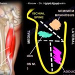
Anatomy of Quadratus Femoris
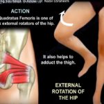
Courtesy: Prof Nabil Ebraheim, University of Toledo, Ohio, USA ANATOMY OF QUADRATUS FEMORIS MUSCLE ORIGIN From the lateral margin of ischial tuberosity INSERTION Into the quadrate tubercle on the intertrochanteric crest between the greater and lesser trochanter ACTION External rotation of the hip and also helps in adduction of thigh NERVE SUPPLY The nerve from […]
Anatomy of Obturator Externus
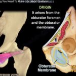
Anatomy of Obturator Externus • The Obturator Externus is one of the six short external rotators of the hip • Origin: It arises from the Obturator foramen and the Obturator membrane. • Insertion: It inserts into the trochanteric fossa of the femur. • Action: it laterally rotates and adducts the thigh • Innervation: The Obturator […]
Anatomy of Flexor Carpi Ulnaris
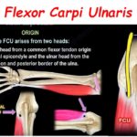
Courtesy: Prof Nabil Ebraheim, University of Toledo, Ohio Anatomy of Flexor Carpi Ulnaris Origin The FCU arises from 2 heads: The humeral head from a common flexor tendon origin of the medial epicondyle and the ulnar head from the olecranon and posterior border of the ulna. Insertion : It inserts on the pisiform bone, the […]
Extensor Digiti Minimi
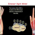
Courtesy: Prof Nabil Ebraheim, University of Toledo, Ohio, USA ANATOMY OF EXTENSOR DIGITI MINIMI It is the sole extensor of the fifth digit ORIGIN From the lateral epicondyle of humerus INSERTION Into the extensor expansion of the fifth digit ACTION Extends the fifth digit at the metacarpophalangeal and interphalangeal joints NERVE SUPPLY Posterior interosseous nerve