
Courtesy: Kaye Wilkins MD, Lynn Staheli MD
www.global-help.org
Supracondylar Fracture of the Humerus in Children
Epidemiology
- Most common elbow fracture in children.
- Peak incidence between 5 and 7 years of age.
- More common in boys.
Reasons for increased incidence
- Maximum ligamentous laxity.
- Weak metaphyseal bone with a thin cortex during remodeling.
- Fall on an outstretched hand is the common mechanism.
Mechanism of Injury
Extension type
- Accounts for approximately 95 to 98 percent of cases.
- Caused by a fall on an extended elbow resulting in hyperextension.
- Failure begins on the anterior cortex under tension.
Flexion type
- Rare.
- Caused by a fall on a flexed elbow.
Types
Based on displacement
- Extension type.
- Flexion type.
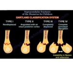
Gartland Classification (Extension Type)
Type I
- Undisplaced fracture.
- Minimal or acceptable displacement.
- Anterior humeral line passes through the capitellum.
Type II
- Angulated fracture with an intact posterior cortex.
- Posterior cortex acts as a hinge.
- Length is maintained.
Type III
- Completely displaced fracture.
- No cortical contact.
- Loss of length with angulation.
- Posteromedial subtype.
- Posterolateral subtype.
Type IV
- Multidirectional instability.
- Unstable in both flexion and extension.
- Less commonly used in routine clinical practice.
Radiological Evaluation
Anterior humeral line
- Should pass through the capitellum.
- Failure to intersect the capitellum indicates unacceptable sagittal angulation.
Fat pad sign
- Posterior fat pad is always abnormal.
- Indicates an occult elbow fracture until proven otherwise.
Baumann angle
- Assesses coronal alignment.
Crescent (half moon) sign
- Suggests varus malalignment.
Acceptable Deformity
- Sagittal angulation of approximately 20 degrees may remodel.
- Coronal plane deformity, particularly varus, does not remodel reliably.
Treatment
Type I fractures
- Above elbow cast.
- Elbow flexed to approximately 90 degrees.
- Immobilization for about 3 weeks.
Pitfall
- Missed medial column collapse may result in cubitus varus.
Type II fractures
Reduction
- Closed reduction under general anesthesia.
- Correct coronal plane deformity first.
- Correct sagittal plane deformity next.
Immobilization
- Stable fractures may be treated with casting.
- Unstable fractures require percutaneous pin fixation.
- Current practice increasingly favors pin fixation.
Type III fractures
- Closed reduction and percutaneous pinning is the treatment of choice.
Reduction principles
- Restore limb length.
- Correct angular deformity.
- Maintain reduction with elbow flexion.
Pin Configuration
Cross pinning
- Medial and lateral K wires.
- Provides maximum mechanical stability.
- Higher risk of iatrogenic ulnar nerve injury.
Lateral pinning
- Two or three lateral K wires.
- Lower risk of ulnar nerve injury.
- Slightly less rotational stability.
- Three lateral pins provide stability comparable to crossed pins in most fractures.
Posteromedial versus Posterolateral Displacement
Posterolateral displacement
- Increased risk of brachial artery injury.
- Increased risk of median nerve and anterior interosseous nerve injury.
- May become irreducible because of brachialis muscle interposition.
Posteromedial displacement
- Increased risk of radial nerve injury.
Indications for Open Reduction
- Irreducible fracture.
- Puckering sign indicating soft tissue interposition.
- Vascular compromise.
- Nerve entrapment.
Surgical approach
Posterolateral displacement
- Anteromedial approach.
Posteromedial displacement
- Anterolateral approach.
Flexion Type Fractures
Features
- Distal fragment displaced anteriorly.
- Tendency to develop valgus deformity.
- Frequently associated with ulnar nerve injury.
Treatment
Type I
- Cast immobilization.
Type II
- Closed reduction.
Type III
- Open reduction is frequently required.
Complications
Early complications
- Median nerve and anterior interosseous nerve injury.
- Radial nerve injury.
- Ulnar nerve injury in flexion type fractures.
- Compartment syndrome.
- Volkmann ischemic contracture.
Late complications
- Cubitus varus due to malunion.
- Elbow stiffness.
- Myositis ossificans, particularly after delayed surgery.
Exam Pearls
- A posterior fat pad sign indicates an occult fracture until proven otherwise.
- Cubitus varus results from malunion rather than growth disturbance.
- Type II fractures are increasingly managed with percutaneous pin fixation.
- Flexion type fractures have a higher rate of open reduction.
- A delay of 6 to 8 hours before surgery is acceptable in the absence of vascular compromise.
Healing
- Fracture union usually occurs within 3 weeks.
- Full range of elbow motion is generally regained within 4 to 6 weeks.

Excellent topics