
Courtesy: Prof Nabile Ebrahiem, University of Toledo, Ohio, USA
Supracondylar Fracture of the Humerus in Children
Introduction
- Most common elbow fracture in children, accounting for approximately 50 percent of pediatric elbow fractures.
- Occurs through the supracondylar region, the weakest part of the distal humerus.
Types of Fracture
Extension type
- Accounts for approximately 95 percent of cases.
- Caused by a fall on an outstretched hand.
- Distal fragment is displaced posteriorly.
Flexion type
- Uncommon.
- Caused by a fall on a flexed elbow.
- Distal fragment is displaced anteriorly.
Nerve Injuries
Extension type
- Anterior interosseous nerve injury is the most common.
- Inability to make the “OK” sign.
- Weakness of the flexor pollicis longus.
- Weakness of the flexor digitorum profundus to the index finger.
- Radial nerve injury is the second most common.
- Wrist and finger extension weakness may be present.
Flexion type
- Ulnar nerve injury is the most common.
- Sensory loss over the little finger.
- Weakness of the intrinsic muscles of the hand.
- Clawing may develop.
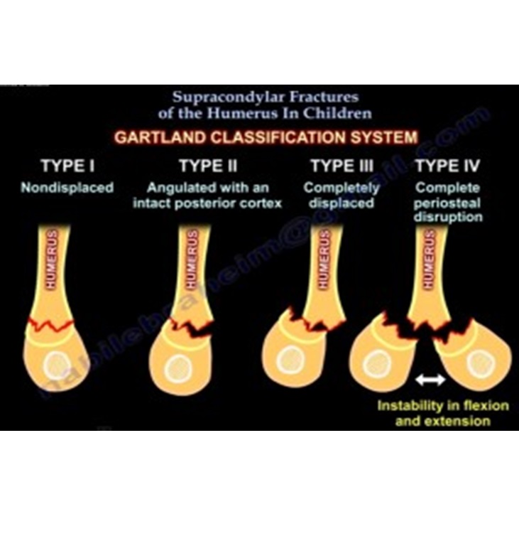
Gartland Classification
Type I
- Undisplaced fracture.
Type II
- Angulated fracture with an intact posterior cortex.
Type III
- Completely displaced fracture.
Type IV
- Multidirectional instability due to complete periosteal disruption.
Radiological Evaluation
Fat pad sign
- Posterior fat pad is always abnormal.
- Suggests an occult elbow fracture.
Anterior humeral line
- Should pass through the middle third of the capitellum.
- In extension type fractures, the capitellum lies posterior to the line.
Baumann angle
- Assesses coronal alignment.
- A decreased Baumann angle indicates an increased risk of cubitus varus deformity.
Clinical Examination
Neurovascular assessment
Anterior interosseous nerve
- Assess with the “OK” sign.
Radial nerve
- Assess wrist and finger extension.
Ulnar nerve
- Assess sensation over the little finger.
- Assess intrinsic hand muscle function.
Treatment
Nonoperative treatment
Indications
- Gartland Type I fractures.
Management
- Above elbow cast or splint.
- Immobilization for 3 to 4 weeks.
- Avoid elbow flexion greater than 90 degrees to prevent vascular compromise.
Operative treatment
Indications
- Gartland Type II fractures.
- Gartland Type III fractures.
- Associated neurovascular compromise.
Technique
- Closed reduction followed by percutaneous pinning.
- Elbow flexion is used to maintain reduction.
- Forearm pronation helps correct varus displacement.
- Fixation with two or three divergent lateral K wires.
- Cross pinning provides greater stability but carries a higher risk of ulnar nerve injury.
Open reduction
Indications
- Failed closed reduction.
- Vascular injury.
- Entrapped soft tissues such as the brachialis muscle.
Surgical principle
- Avoid extensive posterior dissection to preserve blood supply.
Complications
Early complications
- Neuropraxia.
- Compartment syndrome.
- Vascular injury.
Late complications
- Cubitus varus (gunstock deformity) caused by malunion.
- Volkmann ischemic contracture secondary to brachial artery compromise.
- Increased risk with hyperflexion casting beyond 90 degrees.
Pulseless Supracondylar Fracture
Pink pulseless hand
- Hand remains warm and well perfused.
- Treat with closed reduction and percutaneous pinning.
- Observe with serial neurovascular examinations.
Pale pulseless hand
- Hand is cold with poor perfusion.
- Perform urgent closed reduction and percutaneous pinning.
- Proceed to vascular exploration if perfusion does not improve.
Exam Pearls
- The anterior interosseous nerve is the most commonly injured nerve.
- Cubitus varus is the most common late deformity.
- A posterior fat pad sign indicates an occult fracture until proven otherwise.
- Do not immobilize the elbow in more than 90 degrees of flexion.
- Closed reduction and percutaneous pinning is the treatment of choice for displaced supracondylar fractures.
Very nice
Excellent video.
Its a good n covers all issues in schumerus fracture thank u