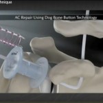
Courtesy: Jayanth Sampath, Ashok Shyam, IORG, OrthoTV
Optimal management of femoral neck fractures in the Elderly
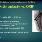
Courtesy: Matthew Lorei, MD, Associate Professor of Orthopaedic Surgery, Temple University Saqib RehmanMD, Director of Orthopaedic Trauma From the 9th Annual Philadelphia Orthopaedic Trauma Symposium June 9, 2017, Lewis Katz School of Medicine at Temple University, Philadelphia
The struggle with diabetes, steroids, and vasculopathy in Ankle Fractures
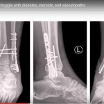
Courtesy: Saqib Rehman MD Director of Orthopaedic Trauma Temple Unviersity Philadelphia USA
Calcium Homeostasis for the FRCS Orth
Courtesy: Quen Tang, FRCS Orth Calcium homeostasis refers to the regulation of serum calcium levels within a narrow physiological range, which is essential for bone health, neuromuscular function, blood coagulation, and cellular signaling. Hormones Involved in Calcium Homeostasis Three major hormones regulate calcium balance: Parathyroid hormone Vitamin D Calcitonin Target Organs of Calcium-Regulating Hormones These […]
Adhesive Arachnoiditis
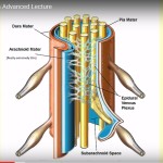
Courtesy: Douglas Gillard, BS, DC, Spine Researcher http://chirogeek.com/
Viscoelastic Properties of Bone
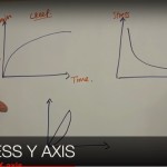
Courtesy: Mock FRCS Cardiff, Saqib Masud FRCS, John Davies FRCS
Understanding the Stress and Strain Curve
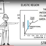
Courtesy: Harry Benjamin Laing MRCS, Ortho M8, FRCS(Tr and Orth Tutorials)
Salenius and Vankka Graph
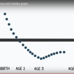
Courtesy:Harry Benjamin Laing, MRCS Ortho M8 FRCS(Tr and Orth) Tutorials https://www.ncbi.nlm.nih.gov/pubmed/1112851
Margins in Tumour Resection

Courtesy: Harry Benjamin Laing Ortho M8 FRCS(Tr and Orth) tutorials
Case discussions on Hand and Wrist Trauma
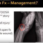
Courtesy: Saqib Rehman Director of Orthopaedic Trauma Temple University Philadelphia USA
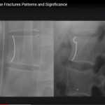
Case discussions on Distal Tibia and ankle fractures
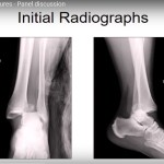
Courtesy: Saqib Rehman Director of Orthopaedic Trauma Temple University Philadelphia USA
Panel Discussion on Post traumatic deformity and Nonunion
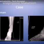
Courtesy: Saqib Rehman MD Director of Orthopaedic Trauma Temple University Philadelphia Pennsylvania USA
Decision making in Humeral shaft fractures
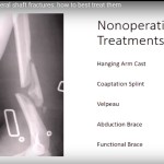
Courtesy: Saqib Rehman MD Director of Orthopaedic Trauma Temple University Philadelphia Pennsylvania USA
Anterior Sternoclavicular Joint Dislocation
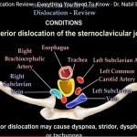
Classification and Management Anatomy of the Sternoclavicular Joint The sternoclavicular joint is a saddle-shaped synovial joint. It is formed by the articulation between: Medial end of the clavicle Manubrium of the sternum First costal cartilage Stabilizing Structures Key stabilizing structures of the sternoclavicular joint include: Anterior and posterior joint capsule Intra-articular fibrocartilaginous disk Interclavicular ligament […]
MRI Anatomy of the Shoulder
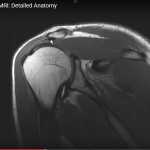
Courtesy: Dr Jean Jose MD, Associate Chief, Musculoskeletal Radiology Section, Associate Professor of Clinical Radiology, University of Miami School of Medicine, Florida, USA MRI Anatomy of the Shoulder Overview The shoulder is a highly mobile and complex joint composed of: Scapula Clavicle Humerus Clinical Importance Understanding shoulder MRI anatomy is essential for evaluating: Rotator […]