Courtesy: Dr Jean Jose MD, Associate Chief, Musculoskeletal Radiology Section, Associate Professor of Clinical Radiology, University of Miami School of Medicine, Florida, USA
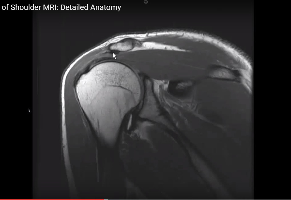
MRI Anatomy of the Shoulder
Overview
The shoulder is a highly mobile and complex joint composed of:
- Scapula
- Clavicle
- Humerus
Clinical Importance
Understanding shoulder MRI anatomy is essential for evaluating:
- Rotator cuff pathology
- Labral injuries
- Ligament injuries
- Neurovascular compression
Key Concept
- Provides wide range of motion
- Stability maintained by soft tissues
Basic Osteology
Scapula
General Features
- Flat triangular bone
- Located on posterior thoracic wall
Important Parts
Body
- Main flat portion
Fossae
- Subscapular fossa (anterior)
- Supraspinous fossa (above spine)
- Infraspinous fossa (below spine)
Borders
- Medial (vertebral) border
- Lateral (axillary) border
Angles
- Superior angle
- Inferior angle
Lateral Structures
- Scapular neck
- Glenoid cavity
- Coracoid process
- Suprascapular notch
Posterior Structures
- Spine of scapula
- Acromion process
Joint
- Acromioclavicular joint
Important Scapular Notches
Suprascapular Notch
- Transmits:
- Suprascapular nerve
- Artery
Spinoglenoid Notch
- Associated with:
- Suprascapular nerve
Proximal Humerus Anatomy
Greater Tuberosity
- Attachments:
- Supraspinatus
- Infraspinatus
- Teres minor
Lesser Tuberosity
- Attachment:
- Subscapularis
Bicipital Groove
- Contains:
- Long head of biceps tendon
Neck
- Anatomical neck
- Surgical neck
Glenoid Labrum
Definition
- Fibrocartilaginous rim around glenoid
Function
- Deepens socket
- Enhances stability
Regions
- Superior
- Inferior
- Anterior
- Posterior
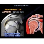
Rotator Cuff
Muscles
- Supraspinatus
- Infraspinatus
- Teres minor
- Subscapularis
Function
- Stabilizes glenohumeral joint
Tendon Insertions
- Supraspinatus – superior facet
- Infraspinatus – middle facet
- Teres minor – inferior facet
- Subscapularis – lesser tuberosity
Important Shoulder Spaces
Triangular Space
Boundaries
- Superior: Teres minor
- Inferior: Teres major
- Lateral: Long head of triceps
Contents
- Circumflex scapular vessels
Triangular Interval
Boundaries
- Superior: Teres major
- Medial: Long head of triceps
- Lateral: Lateral head of triceps
Contents
- Radial nerve
- Profunda brachii artery
Quadrangular Space
Boundaries
- Superior: Teres minor
- Inferior: Teres major
- Medial: Long head of triceps
- Lateral: Surgical neck
Contents
- Axillary nerve
- Posterior circumflex humeral artery
Anterior Shoulder Muscles
Pectoralis Major
- Parts:
- Clavicular
- Sternal
- Abdominal
Insertion
- Lateral lip of bicipital groove
Deltoid Muscle
- Parts:
- Anterior
- Middle
- Posterior
Function
- Abduction and stabilization
Other Muscles
- Pectoralis minor
- Subclavius
Coracoid Process Attachments
Muscles
- Pectoralis minor
- Coracobrachialis
- Short head of biceps
Deltopectoral Groove
Location
- Between deltoid and pectoralis major
Contents
- Cephalic vein
- Thoracoacromial artery
Shoulder Ligaments
Coracoclavicular Ligaments
- Conoid (medial)
- Trapezoid (lateral)
Acromioclavicular Ligaments
- Superior
- Inferior
Coracoacromial Ligament
Function
- Forms coracoacromial arch
Clinical Importance
- Thickening — impingement syndrome
Glenohumeral Ligaments
Types
- Superior
- Middle
- Inferior
Inferior Ligament Components
- Anterior band
- Posterior band
- Axillary pouch
Rotator Cuff Interval
Location
- Between:
- Supraspinatus
- Subscapularis
Contents
- Long head of biceps
- Coracohumeral ligament
- Superior glenohumeral ligament
Biceps Pulley System
Function
- Stabilizes long head of biceps tendon
Components
- Coracohumeral ligament
- Superior glenohumeral ligament
Neurovascular Structures
Suprascapular Nerve
Course
- Through suprascapular notch — spinoglenoid notch
Clinical Importance
- Lesion at notch:
- Affects supraspinatus + infraspinatus
- Lesion at spinoglenoid:
- Affects infraspinatus only
Axillary Nerve
Course
- Passes through quadrangular space
Accompanied by
- Posterior circumflex humeral artery
Summary
Key Structures
- Bones:
- Scapula, clavicle, humerus
- Rotator cuff muscles
- Glenoid labrum
- Shoulder ligaments
- Neurovascular structures
Key Concepts
- Stability mainly from soft tissues
- Rotator cuff is critical stabilizer
- Important spaces:
- Triangular space
- Quadrangular space
- Triangular interval
Thank you I am orthopedic surgeon I want follow any new guidelines