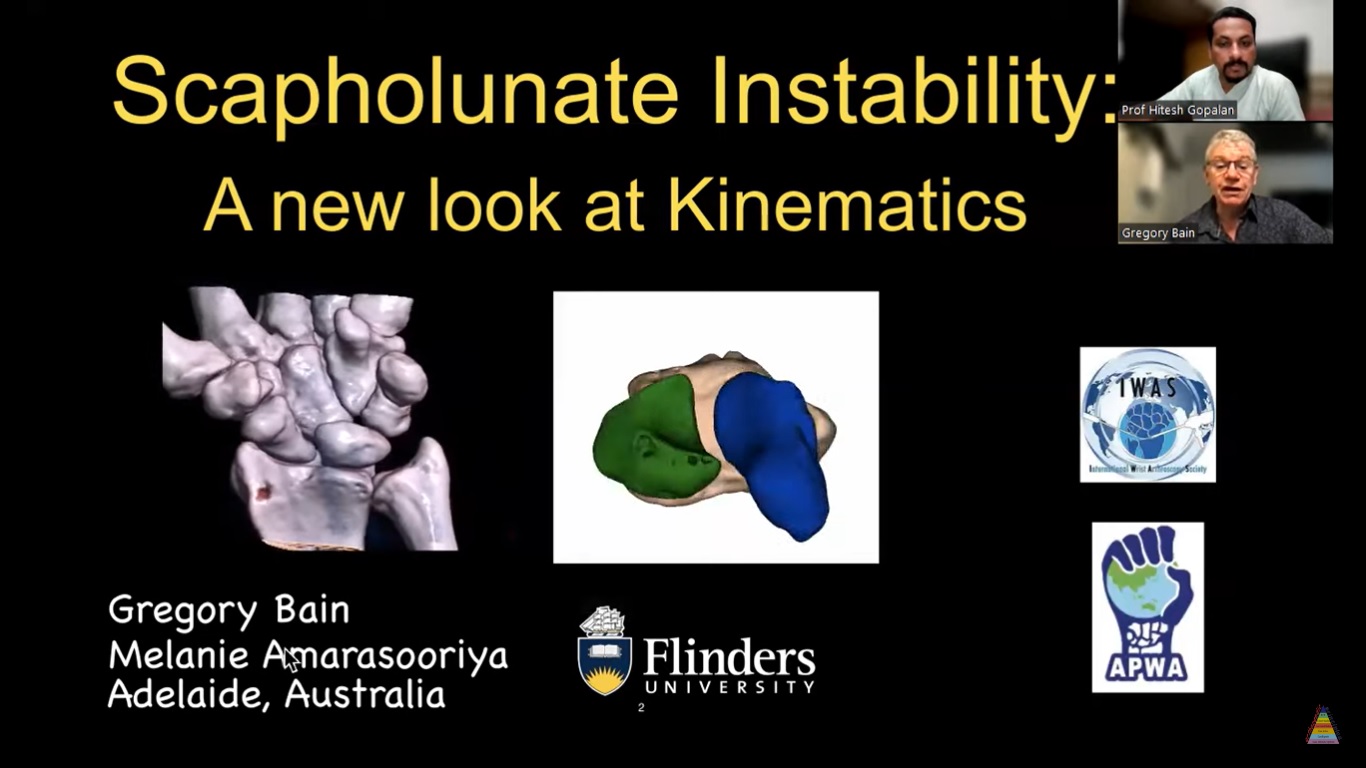
Courtesy: Prof Greg Bain, FRACS, Flinders University, Adelaide, Australia
Scapholunate Instability: Concepts of Scaffolding and Stability
Based on educational concepts in wrist biomechanics and ligament stability
Introduction
Scapholunate instability is one of the most important causes of carpal instability and wrist dysfunction.
Modern understanding of wrist biomechanics has evolved significantly over the past 10–15 years with the use of:
- 4D CT scans for dynamic wrist kinematics
- CT-based wrist models for biomechanical visualization and research
The concept of “scaffolding and stability” emphasizes that wrist stability depends on coordinated ligament complexes rather than isolated ligaments.
Concept of Ligament Complexes
Wrist stability is best understood as interaction between multiple ligament complexes.
Important stabilizing complexes include:
- Dorsal scapholunate complex
- Volar radiolunate complex
- Scaphotrapeziotrapezoid (STT) joint complex
Different ligament groups stabilize different functional zones of the wrist.
Injury may involve:
- A single ligament complex
- Multiple stabilizing complexes simultaneously
Normal Wrist Kinematics
In a normal wrist:
- The scaphoid and lunate move synchronously
- Carpal motion is coordinated and adaptive
- Load is shared efficiently across the carpus
This synchronized movement maintains:
- Stability
- Smooth motion
- Functional load transmission
Pathomechanics of Scapholunate Instability
Basic Problem
Scapholunate instability occurs when coordinated motion between the scaphoid and lunate is lost.
This produces:
- Dissociation of the proximal carpal row
- Abnormal intercarpal motion
- Mechanical instability
Typical Biomechanical Findings
Dorsal Structures
- Dorsal scapholunate ligament complex becomes disrupted
Distal Carpal Row
- STT complex often remains stable
Volar Structures
- Radiolunate and distal radioulnar joint stabilizers are frequently preserved
Resulting Biomechanics
Two relatively stable carpal blocks act across an unstable interval, leading to:
- Deforming forces
- Abnormal motion
- Progressive instability
Dorsal Ligament Complex
The dorsal ligament complex includes:
- Scapholunate ligament
- Dorsal intercarpal ligament (DIC)
- Additional distal stabilizers such as the triquetroscaphoid ligament
This dorsal complex acts like a stabilizing belt across the wrist.
It functions together with the volar ligamentous structures to maintain carpal stability.
Role of the Triquetrum
The triquetrum acts as an important proprioceptive and sensory hub within the wrist.
It receives input from:
- Extensor Carpi Ulnaris (ECU)
- Flexor Carpi Ulnaris (FCU)
- Pisiform-associated structures
Functions include:
- Coordination of carpal motion
- Proprioceptive feedback
- Dynamic stabilization
Biomechanical Changes in Instability
Scaphoid Changes
The scaphoid tends to:
- Flex
- Sublux dorsally over the radius
Lunate Changes
The lunate typically:
- Extends dorsally
- Develops DISI deformity (Dorsal Intercalated Segment Instability)
- Demonstrates reduced motion arc
Clinical Features
Patients may experience:
- Clicking
- Catching
- Pain
- Reduced wrist range of motion
Primary Pathology
The principal problem in scapholunate instability is:
- Radioscaphoid instability
Important concept:
- Scaphoid instability usually occurs before lunate instability
Advanced untreated instability may progress to:
- Carpal collapse
- Degenerative arthritis
- Chronic wrist dysfunction
Capsular and Ligament Injury
Typical injury pattern includes:
- Intrinsic ligament tear
- Extrinsic capsular or ligamentous avulsion
Important healing structures include:
- Capsule
- Ligaments
- Periosteum
Principles of Healing
Importance of Periosteum
Periosteum is considered one of the strongest healing tissues in these injuries.
Synovitis
Inflamed synovial tissue should be debrided because persistent synovitis interferes with healing.
Goal of Repair
The main objective is restoration of:
- Ligament-to-bone attachment
- Capsuloperiosteal continuity
- Stable carpal mechanics
Surgical Techniques
1. Arthroscopic Capsular Plication
Example
Mathoulin technique
Indications
Most useful in:
- Geissler grade 1–3 injuries
Advantages
- Minimally invasive
- Preserves soft tissue
- Avoids complications of open surgery
- Converts symptomatic instability into functional stability
2. Expanded Capsular Repairs
These repairs incorporate a larger portion of the dorsal capsule to improve:
- Global carpal stability
- Force distribution
Arthroscopic techniques are increasingly favored.
3. Window Technique
Small dorsal exposures allow:
- Targeted repair
- Suture anchor placement
- Limited soft tissue disruption
Useful when full arthroscopy is not feasible.
4. Suture Anchor Repair
Technique
- Anchors placed into the scaphoid and lunate
- Capsuloligamentous tissues reattached anatomically
Important Principles
- Prefer cortical fixation
- Respect native ligament footprint
- Utilize tension-band concepts
5. Docking Technique
Anchors are placed into both bones, and sutures are tightened to:
- Reduce the scapholunate interval
- Restore ligament tension
This improves stability by decreasing interbone separation.
Limitations of Tendon Graft Reconstruction
Tendon graft reconstructions have several disadvantages:
- High complication rates
- Persistent scapholunate diastasis
- Inability to restore normal wrist kinematics
- Tendon necrosis
Current evidence suggests tendon grafts do not reliably maintain long-term reduction.
Lessons from ACL Reconstruction
Ligaments possess:
- Broad footprints
- Multiple functional bundles
Joint motion also involves variable centers of rotation.
Therefore:
- A single tendon graft cannot replicate native ligament biomechanics accurately
Problems with Carpal Bone Drilling
Carpal bones possess:
- Thin cortical bone
- Fragile cancellous architecture
Risks of transosseous drilling include:
- Avascular necrosis
- Bone fragmentation
- Disruption of vascular supply
Important point:
- Ligament attachments are superficial, usually less than 1 mm deep
Deep drilling is therefore considered biomechanically unfavorable.
Issues with Synthetic Materials
Synthetic sutures and polyethylene materials may produce:
- Micromotion
- Bone erosion
- Synovitis
- Osteolysis
- Cartilage injury
Because of these issues, synthetic intra-articular constructs are less desirable.
Vascular Considerations
Blood supply to the carpal bones depends on:
- Extraosseous circulation
- Intraosseous circulation
Drilling may disrupt:
- Arterial inflow
- Venous outflow
This increases risk of avascular necrosis of:
- Scaphoid
- Lunate
Denervation
Denervation is generally not recommended during reconstructive procedures because it removes:
- Proprioception
- Neuromuscular feedback
It may be considered only in salvage situations such as:
- Partial wrist fusion
Role of Wrist Models and 3D Printing
Three-dimensional wrist models can reproduce:
- Anatomy
- Ligament relationships
- Carpal mechanics
Applications include:
- Surgical training
- Arthroscopy simulation
- Fracture fixation practice
These models provide a safe and repeatable educational environment.
Clinical Decision-Making
Distal Radius Fracture with Scapholunate Injury
After distal radius fixation:
- Stability should be assessed under fluoroscopy
Management depends on severity:
- Mild instability may be observed
- Significant instability in young or high-demand patients may require arthroscopy and repair
Treatment According to Severity
Grade 1–3 Injuries
- Arthroscopic capsular repair
Advanced but Reducible Injuries
- Window technique with anchor repair
Irreducible or Degenerative Cases
Salvage procedures include:
- Partial wrist fusion
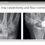
- Proximal row carpectomy
Key Takeaways
- Scapholunate instability is primarily a radioscaphoid instability problem.
- The dorsal ligament complex is essential for wrist stability.
- Tendon graft reconstructions have significant limitations.
- Excessive transosseous drilling should be avoided.
- Capsular-based and minimally invasive repairs are increasingly preferred.
- Successful reconstruction requires respect for:
- Ligament footprint
- Bone biology
- Vascular preservation
Related Posts
 Scapholunate Instability: Evidence-based Management
Courtesy: Dr. Ahlam Arnaout, MD, Paris France Key Insight Despite extensive research, there is no…
- Scapholunate Injuries
Courtesy: Terrence Jose Jerome, Editor, Journal of Hand and Microsurgery
 Scapholunate Ligament Injuries
Courtesy: Vaikunthan Rajarathnam, Hand Surgeon Scapholunate Ligament Injury Introduction The scapholunate ligament is the…
Leave a Reply