Courtesy: William Mackenzie, duPont Hospital, Jefferson Medical College, USA
Definition
• Post-traumatic deformity of distal humerus
• Characterized by:
o Varus angulation
o Extension
o Internal rotation
Also called “Gunstock deformity”
________________________________________
Etiology (Most Important )
• Malunion after supracondylar fracture
o Especially extension type
o Most commonly with posteromedial displacement
Why it happens:
• Inadequate reduction
• Loss of reduction
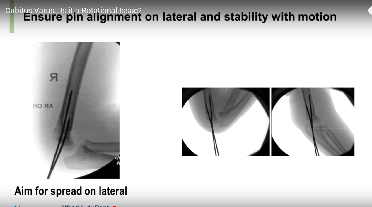
• Poor fixation (casting > pinning risk)
Incidence:
• ~8% with casting
• ~2% with pin fixation
________________________________________
Pathoanatomy
• Primary deformity = Varus tilt (coronal plane)
• NOT mainly due to:
o Translation
o Rotation (minor role only)
________________________________________
Clinical Features
• Cosmetic deformity (main complaint)
• Decreased carrying angle
• Elbow:
o Hyperextension
o Reduced flexion arc (shifted into extension)
________________________________________
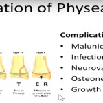
Functional & Late Complications
• Posterolateral rotatory instability (PLRI)
• Tardy ulnar nerve palsy
• Predisposition to:
o Lateral condyle fractures
• Cosmetic concern (major indication for surgery)
________________________________________
Evaluation
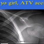
1. X-ray
• Compare both sides
2. Important Measurements
• Baumann’s angle
o Compare with opposite side (more reliable)
• Anterior humeral line
o Should pass through middle 1/3 of capitellum
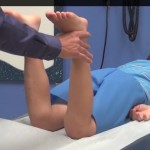
3. Long arm view
• Better for carrying angle assessment
________________________________________
Causes (Less Common)
• Medial growth arrest
• Lateral overgrowth (hyperemia)
• Trochlear osteonecrosis
________________________________________
Treatment
1. Observation
• Most cases (especially mild deformity)
• No functional limitation no surgery
________________________________________
2. Hemiepiphysiodesis
• If progressive deformity in growing child
• Example:
o Lateral growth modulation (8-plate)
________________________________________
3. Corrective Osteotomy (Main Surgical Option)
Gold standard:
Lateral closing wedge osteotomy
________________________________________
Principles of Osteotomy
• Correct:
o Varus
o Extension
o Internal rotation (if needed)
• Avoid lateral prominence
________________________________________
Types of Osteotomy
• Lateral closing wedge (most common )
• Dome osteotomy
• Step-cut osteotomy
• Ilizarov correction (gradual)
________________________________________
Complications of Surgery
• Stiffness
• Nerve injury
• Recurrence
• Lateral condyle prominence
________________________________________
Surgical Tip (High Yield)
• Make wedge distally directed
Prevents lateral bump deformity
________________________________________
Key Exam Pearls
• Most common cause = malunion, NOT growth arrest
• Posteromedial displacement ? varus deformity
• Varus tilt = main deformity
• Cosmetic problem > functional problem
• Compare Baumann angle with opposite side
• Treatment of choice (if needed) – lateral closing wedge osteotomy
Leave a Reply