
Courtesy: Kaye Wilkins MD, Prof Lynn Staheli MD
Sternoclavicular Injuries
Key concept
- Usually represent Salter Harris Type I or II physeal injuries of the medial clavicle rather than true sternoclavicular dislocations.
- Medial clavicular physis is the last epiphysis to ossify and fuse at 20 to 25 years.
- Ligaments are stronger than the physis, so failure occurs through the physis.
Pathology
- Injury occurs through the physis.
- Epiphysis remains in place.
- Metaphysis displaces.
- Intact periosteum allows excellent healing.
Mechanism
- Direct blow to the shoulder.
- Anterior force causes anterior displacement.
- Posterior force causes posterior displacement.
Clinical features
Anterior displacement
- Visible swelling and prominence.
- Local pain.
- Usually benign.
Posterior displacement
- Subtle swelling or depression.
- Dyspnea.
- Dysphagia.
- Venous congestion.
- Facial swelling.
- Risk of mediastinal compression.
Imaging
- Standard radiographs have limited value.
- Serendipity view uses a 40 degree cephalad beam.
- CT scan is the investigation of choice.
Treatment
Anterior displacement
- Usually treated conservatively.
- Closed reduction if required.
- Sling for immobilization.
Posterior displacement
- Orthopedic emergency.
- Urgent closed reduction under general anesthesia.
- Towel clip traction may be required.
- Open reduction if unstable.
Late asymptomatic injuries
- Observation is usually sufficient because remodeling is excellent.
Clavicle Shaft Fractures
General principles
- Very common in children.
- Excellent healing potential.
- Nonunion is rare.
Age related pattern
- Infants usually have minimal displacement.
- Children 1 to 4 years commonly sustain greenstick fractures.
- Children older than 5 years usually have complete displaced fractures.
Treatment
- Sling is the preferred treatment.
- Figure of eight brace is generally unnecessary.
Healing
- Week 1: Pain and deformity are prominent.
- Week 2: Pain decreases.
- Week 3: Callus forms and activity improves.
Indications for surgery
- Open fracture.
- Skin tenting.
- Neurovascular injury.
- Floating shoulder.
- Symptomatic nonunion.
Fixation options
- Plate and screws.
- Elastic intramedullary nail.
Distal Clavicle Injuries
Key concept
- Represent periosteal sleeve injuries rather than true acromioclavicular dislocations.
Pathology
- Periosteum remains intact.
- Coracoclavicular ligaments remain attached.
- Medial clavicle displaces.
- New clavicle forms within the periosteal sleeve.
Treatment
- Type I to III injuries are treated with a sling.
- Type IV to VI injuries usually require surgery.
- Selected symptomatic athletes with Type III injuries may benefit from surgery.
Proximal Humerus Fractures
Key concept
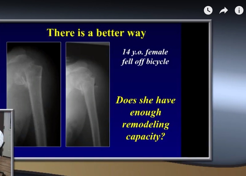
- Excellent remodeling potential.
- Proximal humeral physis contributes about 80 percent of humeral growth.
Muscle forces
Proximal fragment
- Rotator cuff causes abduction and external rotation.
Distal fragment
- Pectoralis major causes adduction and medial displacement.
Age pattern
- Younger children usually sustain metaphyseal fractures.
- Older children more commonly sustain physeal fractures.
Treatment
- Most fractures are treated nonoperatively with a sling.
- Significant deformity remodels well in younger children.
Indications for surgery
- Open fracture.
- Neurovascular injury.
- Failed closed reduction.
- Older child with severe displacement.
- High demand athlete.
Fixation options
- Percutaneous Kirschner wires.
- Retrograde elastic intramedullary nails.
- Cannulated screws in older adolescents.
Complications
- Malunion causing impingement.
- Reduced range of motion.
Metaphyseal Versus Physeal Fractures
Physeal fractures
- Greater rotational deformity.
- Stronger muscle deforming forces.
- Good remodeling.
Metaphyseal fractures
- Less rotational deformity.
- Less muscle displacement.
- Excellent remodeling.
Special Points
Always assess
- Neurovascular status.
- Shoulder dislocation.
- Coracoid fracture.
- Brachial plexus injury.
Floating shoulder
- Combination of clavicle and scapular neck fracture.
- Usually requires stabilization of the clavicle.
Exam Pearls
- Sternoclavicular injury in children is usually a physeal injury rather than a true dislocation.
- Posterior sternoclavicular injury is life threatening.
- Most clavicle fractures are treated nonoperatively.
- Distal clavicle injuries are periosteal sleeve injuries.
- Proximal humerus has the greatest remodeling potential in the body.
- Surgery for proximal humerus fractures is rarely required except in older children with severe displacement.
Leave a Reply