
Courtesy: Kaye Wilkins MD, Prof Lynn Staheli MD www.global-help.org
Pediatric Foot Fractures and Dislocations
Overview
Pediatric foot fractures
- Rare because the immature foot is highly flexible.
- Thick cartilage absorbs impact forces.
- Forces are often transmitted proximally to the ankle, tibia, or knee rather than causing foot fractures.
Minor Foot Fractures
Metatarsal base fractures
- Usually result from trivial trauma.
- Treated with immobilization alone.
- Healing is rapid with excellent outcomes.
Talus Fractures
Mechanism of injury
- Forced dorsiflexion combined with pronation.
- Anterior tibia impacts the talar neck causing fracture.
Imaging
- Canale view is the preferred radiographic view.
- Foot is pronated approximately 15 degrees.
- X ray beam is angled approximately 75 degrees.
- Best demonstrates talar neck alignment and displacement.
Acceptable displacement
- Less than 5 mm displacement.
- Less than 5 degrees angulation.
Reduction principle
- Reverse the injury mechanism.
- Plantarflexion.
- Supination.
Blood supply
- Talus has a retrograde blood supply.
- High risk of avascular necrosis after fracture.
Hawkins classification
- Type I: Undisplaced fracture with 0 to 13 percent risk of avascular necrosis.
- Type II: Subtalar subluxation with 20 to 50 percent risk.
- Type III: Associated ankle dislocation with high risk.
- Type IV: Associated talonavicular dislocation with very high risk.
Hawkins sign
- Subchondral lucency of the talar dome.
- Indicates preserved blood supply.
- Suggests a good prognosis.
Complications
- Avascular necrosis.
- Subtalar arthritis.
- Malunion.
Osteochondral Lesions of the Talus
Clinical features
- May be traumatic or atraumatic.
- Pain.
- Clicking.
- Joint effusion.
Treatment
- Only symptomatic lesions require treatment.
- Arthroscopy.
- Open reduction and internal fixation or bone grafting for large lesions.
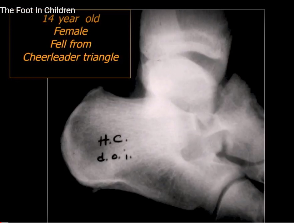
Calcaneal Fractures
Mechanism
- Usually caused by axial compression following a fall from height.
Associated injuries
- Always evaluate for lumbar spine fractures.
Types
Stress fracture
- Common in athletes.
- Pain increases with activity.
- Bone scan is useful early.
- X rays may become positive later.
Extra articular fracture
- Usually treated conservatively.
Intra articular fracture
- Involves the subtalar joint.
- CT scan is recommended.
Bohler angle
- Normal is 20 to 40 degrees.
- Decreased angle indicates calcaneal collapse.
Treatment
- Mild displacement: Conservative treatment.
- Significant displacement: Open reduction and internal fixation or screw and pin fixation.
Special Calcaneal Injury
Nutcracker fracture of the cuboid
Mechanism
- Forefoot abduction compresses the cuboid between the calcaneus and metatarsals.
Treatment
- Restore lateral column length.
- Bone grafting.
- Internal fixation.
Midfoot Injuries
Lisfranc injury
- Involves the base of the second metatarsal with tarsometatarsal disruption.
- Requires anatomical reduction.
- Usually managed with internal fixation.
Metatarsal Fractures
Base of the fifth metatarsal
- Differentiate avulsion fracture from Jones fracture.
Jones fracture
- Occurs at the proximal diaphysis.
- Poor blood supply.
- High risk of delayed union and nonunion.
- Often treated with intramedullary screw fixation.
Metatarsal neck fractures
- Excellent remodeling potential in children.
- Usually managed conservatively.
Phalangeal Fractures
Management
- Commonly caused by crush injuries.
- Buddy taping is sufficient in most cases.
- Surgery is rarely required.
Baseball toe
- Avulsion fracture of the distal phalanx.
- Often requires fixation similar to mallet finger.
Complications
- Avascular necrosis of the talus.
- Subtalar arthritis.
- Growth arrest.
- Malunion.
- Nonunion, especially in Jones fractures.
- Foot compartment syndrome requiring medial and dorsal fasciotomy.
Exam Pearls
- Pediatric foot fractures are uncommon because of the flexibility of the immature foot.
- Talus fracture is the most important pediatric foot fracture because of the risk of avascular necrosis.
- Canale view is the preferred radiographic view for talar neck fractures.
- Hawkins sign indicates preserved talar vascularity.
- Always evaluate the lumbar spine in patients with calcaneal fractures.
- Nutcracker fracture refers to compression fracture of the cuboid.
- Jones fractures have a high risk of delayed union and nonunion.
- Lisfranc injuries usually require anatomical reduction and fixation.
Leave a Reply