
Courtesy: Kaye Wilkins MD
Lynn Staheli MD
www.global-help.org
Unappreciated Fractures Around the Pediatric Elbow
Why “Unappreciated” rather than “Missed”?
- “Missed fracture” may imply negligence.
- “Unappreciated fracture” acknowledges the genuine diagnostic difficulty created by pediatric anatomy.
Why Pediatric Elbow Fractures Are Difficult to Diagnose
1. Children are not miniature adults
- Multiple physes.
- Multiple ossification centers.
- Large cartilaginous components that are not visible on X-ray.
2. Know CRITOE
| Ossification center | Approximate age |
|---|---|
| Capitellum | 1–2 yr |
| Radial head | 3–4 yr |
| Internal/medial epicondyle | 5–7 yr |
| Trochlea | 8–10 yr |
| Olecranon | 9–10 yr |
| External/lateral epicondyle | 10–12 yr |
Key principle: Before an ossification center appears, the corresponding structure is largely cartilage and a fracture may therefore be radiographically occult.
Important Unappreciated Elbow Injuries
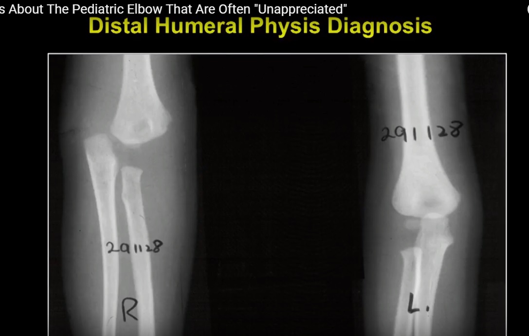
1. Distal Humerus Physeal Separation ?
The source identifies this as the most commonly missed injury.
Features
- Usually in children <7 years.
- The entire distal humeral fragment may displace.
- Typical displacement: posteromedial.
Radiological clue
Radiocapitellar alignment is maintained.
This helps distinguish it from an elbow dislocation.
May mimic
- Elbow dislocation.
- Lateral condyle fracture.
Diagnosis
- Arthrogram — particularly useful.
- MRI is rarely required.
2. Medial Condyle Fracture ?
Often mistaken for a medial epicondyle fracture.
Important clue
In a young child, the trochlea may not yet be ossified, so a substantial cartilaginous fracture component may be invisible.
Rule
“Medial epicondyle fracture before age 8 = medial condyle fracture until proven otherwise.”
Complications
- AVN.
- Deformity.
3. TRASH Lesions
TRASH = The Radiographic Appearance Seemed Harmless
These are injuries with subtle radiographic findings despite potentially significant pathology.
The important lesson is:
A normal-looking X-ray does not necessarily mean a normal pediatric elbow.
Lateral Condyle vs Distal Humerus Physeal Separation
| Feature | Lateral condyle | Distal humerus physis |
|---|---|---|
| Displacement | Lateral | Posteromedial |
| Radiocapitellar alignment | Lost | Maintained |
| Treatment | Often ORIF | Closed reduction may be possible |
Non-Accidental Injury
A particularly important red flag is:
Distal humeral physeal separation in a non-walking child
Consider non-accidental injury and perform appropriate evaluation, including:
- Skeletal survey.
- Search for associated injuries such as rib or skull fractures.
Don’t Stop at the Obvious Injury
Forearm injuries
Image the entire forearm and elbow when appropriate.
Look for associated injuries such as:
- Distal radius fracture + radial neck fracture.
- Monteggia lesion.
Knee pain
Knee pain can originate from the hip.
The source specifically highlights SCFE as an example.
Radial head fracture
Look for associated lateral collateral ligament injury, which can contribute to elbow instability.
AVN of the Trochlea
The source describes two patterns:
Type A
- Partial involvement.
- “Fishtail deformity.”
Type B
- Complete involvement.
- More severe deformity, including cubitus varus/valgus.
Cause
Disruption of the blood supply to the distal humerus.
? Golden Rules
- Clinical examination comes first — don’t rely solely on the X-ray.
- Image the entire relevant limb when an associated injury is possible.
- Know CRITOE.
- Always assess alignment:
- Radiocapitellar line
- Anterior humeral line
- Look actively for associated injuries.
- Consider non-accidental injury in a non-ambulatory child or when the history is inconsistent.
? Ultimate Exam Pearl
“Most unappreciated pediatric fractures are due to failure to understand ossification and incomplete examination—not lack of knowledge.”
Leave a Reply