
Courtesy: Dr. Shital Parikh, Dr Ashok Shyam, Ortho TV
TRASH Elbow Injuries (Pediatric Elbow)
Definition
TRASH = “The Radiographic Appearance Seemed Harmless.”
These are pediatric elbow injuries that:
- Appear normal or trivial on plain X-rays.
- Are actually significant injuries, often intra-articular.
- Are frequently missed initially and may lead to poor outcomes.
Why Are They Easily Missed?
The pediatric elbow has:
- Multiple ossification centers.
- A large cartilaginous component that is not visible on X-ray.
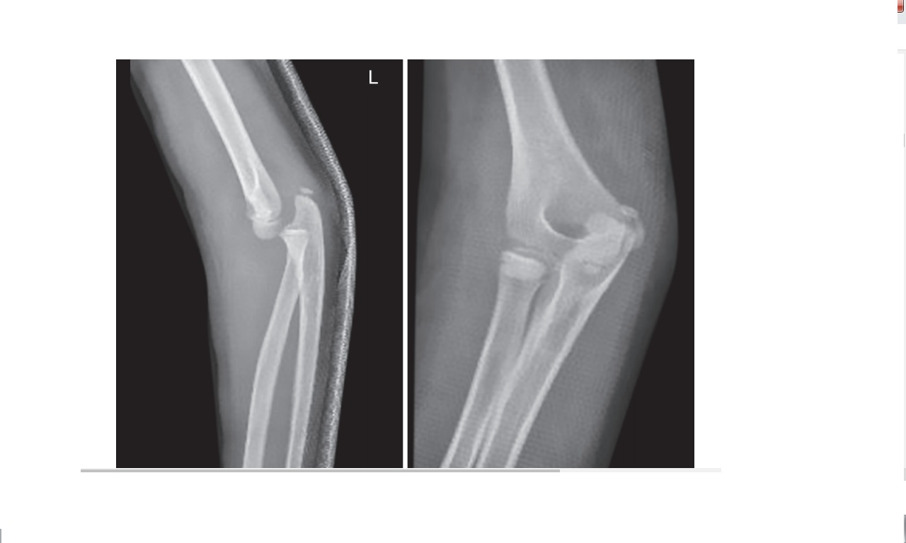
Therefore, a tiny bony “flake” on an X-ray may actually represent a much larger cartilaginous fracture fragment.
Typical Clinical Scenario
- Usually a child younger than 10 years.
- Mechanism: fall.
- Closed, isolated injury.
- Examination may appear nearly normal, with only mild pain or swelling.
This creates a high risk of missed diagnosis.
Common TRASH Injuries
- Occult fractures.
- Shear fractures of the capitellum or trochlea.
- Cartilage injuries involving unossified fragments.
- Radial head fractures.
- Entrapped medial epicondyle fragments.
- Subtle Monteggia variants.
- Transphyseal distal humeral injuries in young children.
Red Flags
Maintain a high index of suspicion when there is:
- An elbow dislocation with a small bony fragment.
- Persistent pain despite a “normal” X-ray.
- Persistent swelling.
- Elbow stiffness.
- Limited range of motion.
- Significant ecchymosis or soft-tissue swelling.
Systematic X-ray Evaluation
1. Fat Pad Signs
Posterior fat pad
- Always abnormal.
- Indicates an intra-articular injury.
Anterior fat pad
- A small anterior fat pad can be normal.
- A prominent “sail sign” suggests an effusion or hemarthrosis.
2. Radiocapitellar Line
- Draw a line along the radial neck.
- It should pass through the capitellum on AP, lateral, and oblique views.
- It is less reliable in children younger than 4 years.
3. Soft-Tissue Shadows
- Asymmetry or swelling may indicate a hidden injury.
- The pattern of swelling can help localize medial versus lateral pathology.
4. Normal Anatomical Clues
- The capitellum is wider posteriorly than anteriorly.
- The normal “teardrop” appearance should be recognized to avoid mistaking anatomy for pathology.
5. Direction of Displacement
This can help distinguish injuries:
Transphyseal distal humerus fracture – Posteromedial
Elbow dislocation – Posterolateral
MRI
Best for:
- Cartilage injuries.
- Occult fractures.
CT
Best for:
- Bony fragments.
Arthrogram
Useful when MRI is unavailable or impractical.
It demonstrates the intra-articular anatomy clearly.
Important Injury Patterns
Radial Head Fractures
- May be radiographically subtle.
- If missed, they may lead to early arthritis and poor outcomes.
Lateral Condyle Fractures
- May extend medially and become unstable.
- Soft-tissue swelling may provide an important clue.
Medial Epicondyle Fracture
- Often associated with elbow dislocation.
- The fragment may become entrapped within the joint and then requires ORIF.
Medial Condyle Fracture
- Rare (less than 1%).
- Has a high complication rate, including AVN and nonunion.
Shear Fractures of Capitellum or Trochlea
- Frequently missed on initial X-rays.
- Usually require open reduction and fixation.
Coronoid Fracture
- May occur with elbow dislocation and radial head injury.
- Produces elbow instability.
Transphyseal Distal Humerus Fracture
- Seen mainly in children younger than 3 years.
- May mimic elbow dislocation.
- The radiocapitellar alignment remains preserved.
Management Principles
- Maintain a high index of suspicion.
- If the clinical findings do not match the X-ray, obtain MRI or an arthrogram.
- Early, accurate diagnosis is essential.
- Intra-articular injuries should be anatomically reduced and fixed with K-wires or screws when indicated.
- Avoid prolonged immobilization to reduce the risk of elbow stiffness.
Complications of Missed TRASH Injuries
- Elbow stiffness (most common).
- Chronic instability.
- Malunion.
- Early arthritis.
- Loss of pronation and supination.
High-Yield Exam Pearls
- TRASH = The Radiographic Appearance Seemed Harmless.
- Small fragment = big problem.
- Posterior fat pad = fracture until proven otherwise.
- Elbow dislocation in a child should always prompt a search for an associated fracture.
- Persistent pain or stiffness after a normal X-ray warrants advanced imaging.
- Early recognition and anatomic treatment prevent long-term disability.
Leave a Reply