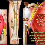
? Courtesy: Prof Nabil Ebraheim, University of Toledo, Ohio, USA RADIAL NERVE It is a major peripheral nerve in the upper limb. It arises from the posterior cord of brachial plexus and contains fibres from nerve roots C5 to T1 COURSE It arises in the axilla posterior to axillary artery Traverses through the posterior […]
-Applied Anatomy
Anatomy of Cubital fossa, Popliteal fossa and the Femoral Triangle
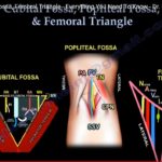
Courtesy: Prof Nabil Ebraheim, University of Toledo, Ohio, USA Overview Certain anatomical regions contain important neurovascular structures arranged in a specific order. Three clinically important regions are: Cubital fossa Popliteal fossa Femoral triangle Knowledge of the contents and arrangement of these structures is essential for clinical examination, surgical procedures, and emergency interventions. Cubital Fossa Location […]
Anatomy of the Femoral Triangle
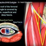
Courtesy: Prof Nabil Ebraheim, University of Toledo, Ohio, USA ANATOMY OFTHE FEMORAL TRIANGLE Femoral triangle is a superficial triangular space located on the anterior aspect of the thigh just inferior to the inguinal ligament . The boundaries of the triangle include the medial border of the sartorius on the lateral aspect ,medial border of adductor […]
Anatomy and Contents of the Popliteal fossa
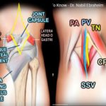
Courtesy: Prof Nabil Ebraheim, University of Toledo, Ohio, USA Popliteal fossa is a shallow depression located at the back of the knee joint , bounded by biceps femoris superolaterally ,semimembranosus and semitendinosus superomedially . Inferior boundary of this space is made by the medial and lateral heads of gastrocnemius muscle . Coming to the base […]
Anatomy and Tributaries of the Anterior Tibial Artery
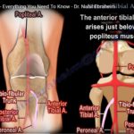
Courtesy: Prof Nabil Ebraheim, University of TOledo, Ohio, USA Overview The anterior tibial artery is one of the terminal branches of the popliteal artery. It supplies the anterior compartment of the leg and continues distally as the dorsalis pedis artery. Understanding its course and anatomical relationships is important in trauma, orthopedic surgery, and vascular assessment. […]
Anatomy and Contents of the Anatomical Snuff Box
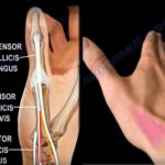
Courtesy: Prof Nabil Ebraheim, University of Toledo, Ohio, USA The anatomical snuffbox is a small triangular depression located on the dorsoradial aspect of the wrist.People used this space to place and sniff the powdered tobacco or “snuff”, hence the name.The base of this triangular space is proximal with the apex pointing towards the thumb.The anatomical […]
Anatomy and Contents of the Cubital Fossa
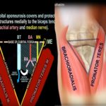
Courtesy: Prof Nabil Ebraheim, University of Toledo, Ohio, USA The cubital fossa is a triangular depression located in front of the anterior elbow.The medial border is formed by the pronator teres which arises from the medial epicondyle of the humerus.The lateral border is formed by the brachioradialis muscle which arises from the lateral supracondylar ridge […]
Tendon arrangement in the Ankle Region
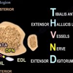
Courtesy: Prof Nabil Ebraheim, University of Toledo, Ohio, USA Tendons at the anterior compartment of ankle are Tibialis anterior,Extensor hallucis longus ,Extensor digitorum longus. Also passing through the anterior compartment is the anterior tibial artery and deep peroneal nerve. The superior and inferior extensor retinaculum overlies this area. The mnemonic to remember arrangement of anterior […]
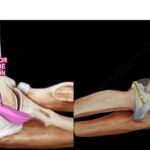
Anatomy Of The Vastus Lateralis Muscle
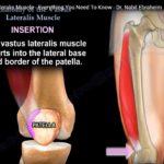
Courtesy: Prof Nabil Ebraheim, University of Toledo, Ohio, USA Video describes the Anatomy of Vastus Lateralis Muscle which is a part of Quadriceps Femoris. Vastus lateralis originates from the greater trochanter of the femur and from the lateral lip of the Linea Aspara. It loops around the shaft of femur from the posterior to anterior […]
Anatomy of Rectus Femoris Muscle
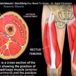
Courtesy: Prof Nabil Ebrhaeim, University of Toledo, Ohio, USA Video describes Anatomy of Rectus Femoris which is the Anterior Muscle of Quadriceps Femoris. Rectus Femoris Originates from the pelvis as two heads. 1: Straight Head From Anterior Inferior Iliac Spine and 2: Reflected head originate from a groove superior to acetabulum. The Muscle is inserted […]
Anatomy Of The Extensor Digitorum Communis
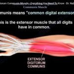
Courtesy: Prof Nabil Ebraheim, University of Toledo, Ohio, USA Video describes Anatomy of Extension Digitorum Communis Muscle. It Originates from the Lateral Epicondyle of Humerus which is the Common Extensor Origin.The muscle is inserted into Extensor expansions of medial four digits.It is supplied by Posterior interosseous nerve (C7).The Primary Action of this muscle is to […]
Anatomy of Superior and Inferior Gemellus
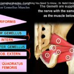
Courtesy: Prof Nabil Ebraheim, University of Toledo, Ohio, USA This Video describes The Anatomy of Superior and Inferior Gemellus Muscle. The Superior Gemellus Muscle originates the Ischial Spine and Inferior Gemellus Muscle originate from the posterior portion of the Ischial tuberosity and the lateral Obturator. Both the Muscles insert into the medial surface of greater […]
Anatomy of the Ligaments of Elbow
Courtesy: Prof Nabil Ebraheim, University of Toledo, Ohio, USA Anatomy of ligaments of elbow joint The elbow joint is a hinge joint, formed by the articulation between the lower end of the humerus with the ulna and head of the radius. Stability of the elbow joint depends upon the inherent stability of the articulating surfaces, […]
Anatomy Of The Coracobrachialis Muscle
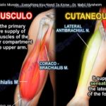
Courtesy: Prof Nabil Ebraheim, University of Toledo, Ohio, USA The video describes The Anatomy of Coracobrachialis Muscle. It Originate from tip of the Coracoid Process. It is inserted into middle third of the medial border of humeral Shaft.The Innervation of the Coracobrachialis Muscle comes from the Musculocutaneous Nerve.The Primary Action of this Muscle is to […]
Anatomy Of The Pectoralis Minor Muscle
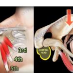
Courtesy: Prof Nabil Ebraheim, University of Toledo, Ohio, USA ANATOMY OF PECTORALIS MINOR Pectoralis minor is a thin triangular muscle in the upper part of chest situated beneath the pectoralis major muscle It joins shoulder girdle to thorax ORIGIN From anterior surface of superior margins of 3 rd 4 th and 5 th ribs near […]