Courtesy: Prof Nabil Ebraheim, University of Toledo, Ohio, USA
Overview
- Certain anatomical regions contain important neurovascular structures arranged in a specific order.
- Three clinically important regions are:
- Cubital fossa
- Popliteal fossa
- Femoral triangle
- Knowledge of the contents and arrangement of these structures is essential for clinical examination, surgical procedures, and emergency interventions.
Cubital Fossa
Location
- The cubital fossa is a triangular depression located on the anterior aspect of the elbow joint.
Important Structures in the Cubital Fossa
The contents are arranged from medial to lateral.
- Median nerve
- Brachial artery and its bifurcation into:
- Radial artery
- Ulnar artery
- Biceps brachii tendon
- Radial nerve and its branches
Biceps Tendon and Aponeurosis
- The biceps brachii tendon lies lateral to the brachial artery.
- It inserts primarily into the radial tuberosity.
- A secondary expansion called the bicipital aponeurosis extends medially.
Functions of the bicipital aponeurosis:
- Covers and protects deeper structures in the cubital fossa.
- Provides protection to:
- Brachial artery
- Median nerve
Radial Nerve
- Located lateral to the biceps tendon.
- Divides into branches including the posterior interosseous nerve.
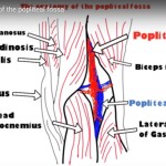
Popliteal Fossa
Location
- The popliteal fossa is a shallow depression located posterior to the knee joint.
Contents of the Popliteal Fossa
The structures are arranged from medial to lateral.
- Popliteal artery
- Popliteal vein
- Small saphenous vein
- Tibial nerve
- Common peroneal nerve
Important point:
- The common peroneal nerve runs along the upper lateral border of the popliteal fossa.
Femoral Triangle
Location
- The femoral triangle is a superficial triangular space located on the anterior aspect of the upper thigh, just inferior to the inguinal ligament.
Contents of the Femoral Triangle
The structures are arranged from lateral to medial.
- Femoral nerve
- Femoral artery
- Femoral vein
Additional contents include:
- Deep inguinal lymph nodes
Key Arrangement
The femoral triangle contents follow the classic lateral-to-medial sequence:
- Nerve
- Artery
- Vein
This arrangement is clinically important during procedures such as:
- Femoral arterial catheterization
- Femoral nerve block
- Vascular access
Summary Points
- The cubital fossa contains the median nerve, brachial artery, biceps tendon, and radial nerve arranged from medial to lateral.
- The popliteal fossa lies behind the knee and contains major vessels and nerves including the popliteal artery, vein, tibial nerve, and common peroneal nerve.
- The femoral triangle lies below the inguinal ligament and contains the femoral nerve, femoral artery, and femoral vein arranged from lateral to medial.


Leave a Reply