
Courtesy: Sameer Qureshi, Consultant Orthopaedic Surgeon, Ujjain, MP BLOOD SUPPLY OF THE LONG BONE BASIC ANATOMY OF THE LONG BONE The elongated central part of the long bone is called the diaphysis. The enlarged area of the bone at the ends is called the epiphysis and the intermediate bone segment between the two is called […]
-Applied Anatomy
Anatomy of Iliopsoas Muscle

Courtesy: Prof Nabil Ebraheim, University of Toledo, Ohio, USA ANATOMY OF ILIOPSOAS MUSCLE INTRODUCTION: IT INCLUDES 3 MUSCLES : PSOAS MAJOR,PSOAS MINOR(if present),ILIACUS. PSOAS MAJOR ORIGIN : It arises from the transverse processes and lateral aspect of vertebral bodies T12 – L5. COURSE :Runs downwards across the pelvic brim and then passes deep to the […]
Anatomy of The Gluteus Maximus Muscle
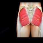
Courtesy: Prof Nabil Ebraheim, University of Toledo, Ohio, USA GLUTEUS MAXIMUS Gluteus maximus is the largest and heaviest muscle in the body. It is the most superficial of all gluteal muscles that are located at the posterior aspect of hip joint. ORIGIN: The gluteus maximus originates from The gluteal surface of ilium Lumbar fascia Sacrum […]
Anatomy Of The Tensor Fascia Lata Muscle
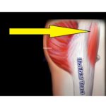
Courtesy: Prof Nabil Ebraheim, University of Toledo, Ohio, USA Tensor fascia lata muscle lies between tendon of gluteus maximus and tensor fascia lata at the middle of upper area of the thigh. Origin – anterior part of outer lip of iliac Crest Insertion – iliotibial tract at middle third of thigh (iliotibial band) Function: Flexion […]
Anatomy Of The Latissimus Dorsi Muscle
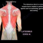
Courtesy: Prof Nabil Ebraheim, University of Toledo, Ohio, USA Anatomy of the Latissimus Dorsi Muscle Latissimus dorsi muscle is anatomically is a muscle on the back but functionally is a muscle on the upper limb. It is the broadest muscle of the back. It is one of the strongest muscle and is used in sports […]
Anatomy of the Trapezius Muscle
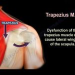
Courtesy: Prof Nabil Ebraheim, University of Toledo, Ohio, USA Anatomy of Trapezius muscle Trapezius is a large superficial muscle that extends from the back of the skull,back of the neck and back of the thorax. Origin: The upper fibres of the trapezius muscle arise from the external occipital protruberance and the medial third of the […]
Anatomy Of The Teres Minor Muscle
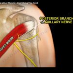
Courtesy: Prof Nabil Ebraheim, University of Toledo, Ohio, USA Anatomy of the teres minor muscle The teres minor is a narrow muscle which originates from the posterior lateral surface of the scapula and inserts into the greater tuberosity of the humerus . Teres minor is innervated by the posterior branch of axillary nerve . The […]
Anatomy Of The Teres Major Muscle
Courtesy: Prof Nabil Ebraheim, University of Toledo, Ohio, USA Anatomy of Teres Major Muscle Origin- dorsal aspect of inferior angle of scapula Insertion- medial lip of intertubercular groove of the humerus One of the muscle that connect scapula to the humerus Teres major muscle does not attach to the capsule of glenohumeral joint Teres major […]
Anatomy and Blood supply of Talus
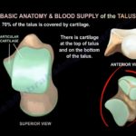
Courtesy: Prof Nabil Ebraheim, University of Toledo, Ohio, USA Basic Anatomy of the Talus Approximately 70% of the talus surface is covered by articular cartilage. Talus consists of head, neck, body, lateral process, and posterior process. Large cartilage coverage contributes to high risk of post?traumatic arthritis after fractures. Blood Supply of the Talus Primary blood […]
Anatomy Of The Popliteus Muscle
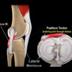
Courtesy: Prof Nabil Ebrhaeim, University of Toledo, Ohio, USA Popliteus muscle is one of the deep flexors of the knee. It is a thin triangular muscle with narrow tendinous origin and broad insertion at the posterior aspect of the knee joint. ORIGIN Arise from the anterior part of the popliteal grove on the lateral surface […]
Anatomy of Plantaris Muscle
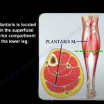
Courtesy: Prof Nabil Ebraheim, University of Toledo, Ohio, USA Anatomy Plantaris muscle arises from the lateral supracondylar ridge of the femur. Origin is located above the lateral head of the gastrocnemius muscle. The tendon passes between the gastrocnemius and soleus muscles. It inserts on the medial side of the calcaneus. It lies in the superficial […]
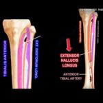
Anatomy Of The Tibialis Anterior Muscle
Courtesy: Prof Nabil Ebraheim Tibialis Anterior Muscle The Tibialis Anterior muscle is located within the anterior extensor compartment of the leg. Origin: From upper half and lateral condyle of the tibia Insertion: into the medial cuneiform and first metatarsal base of the foot Tibialis anterior muscle comes from the lateral surface of the tibia and […]
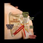
Anatomy Of The Subclavius Muscle
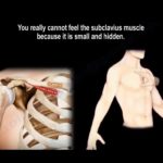
Courtesy: Prof Nabil Ebraheim, University of Toledo, Ohio, USA ANATOMY OF SUBCLAVIUS MUSCLE The subclavius muscle is the short muscle of shoulder girdle. ORIGIN: at the junction of first rib and costal cartilage. INSERTION: groove of subclavius in the inferior surface of the middle third of the clavicle. It lies behind the pectoralis major NERVE […]
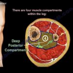
Anatomy Of The Extensor Hallucis Longus Muscle
? Courtesy: Prof Nabil Ebraheim, University of Toledo, Ohio, USA Anatomy of Extensor Hallucis Longus • The extensor hallucis longs muscle lies in the anterior extensor compartment of the leg. • There are four muscle compartments within the leg (Q: “How many muscle compartments are present in the leg”) Anterior Compartment Lateral Compartment Superficial […]
Anatomy Of The Extensor Digitorum Longus Muscle
Courtesy: Prof Nabil Ebraheim, University of Toledo, Ohio, USA Leg Compartments The leg contains four compartments: anterior, lateral, superficial posterior, and deep posterior. The extensor digitorum longus lies in the anterior compartment of the leg. Muscles of the Anterior Compartment Tibialis anterior. Extensor hallucis longus. Extensor digitorum longus. Peroneus tertius. All muscles in the anterior […]