Courtesy: Prof Nabil Ebraheim, University of Toledo, Ohio, USA
Layers of the Articular Cartilage and Cartilage Healing
Introduction: Does Articular Cartilage Heal After Injury?
-
Articular cartilage has a very limited capacity for healing.
-
The ability of cartilage to heal depends on the depth of the injury and whether it reaches the underlying bone.
-
Because articular cartilage is avascular, injuries confined to cartilage alone generally do not heal.
Structural Anatomy of Articular Cartilage
When examining a joint surface in detail, the following structures are identified:
-
Articular cartilage
-
Subchondral bone
-
Cancellous bone
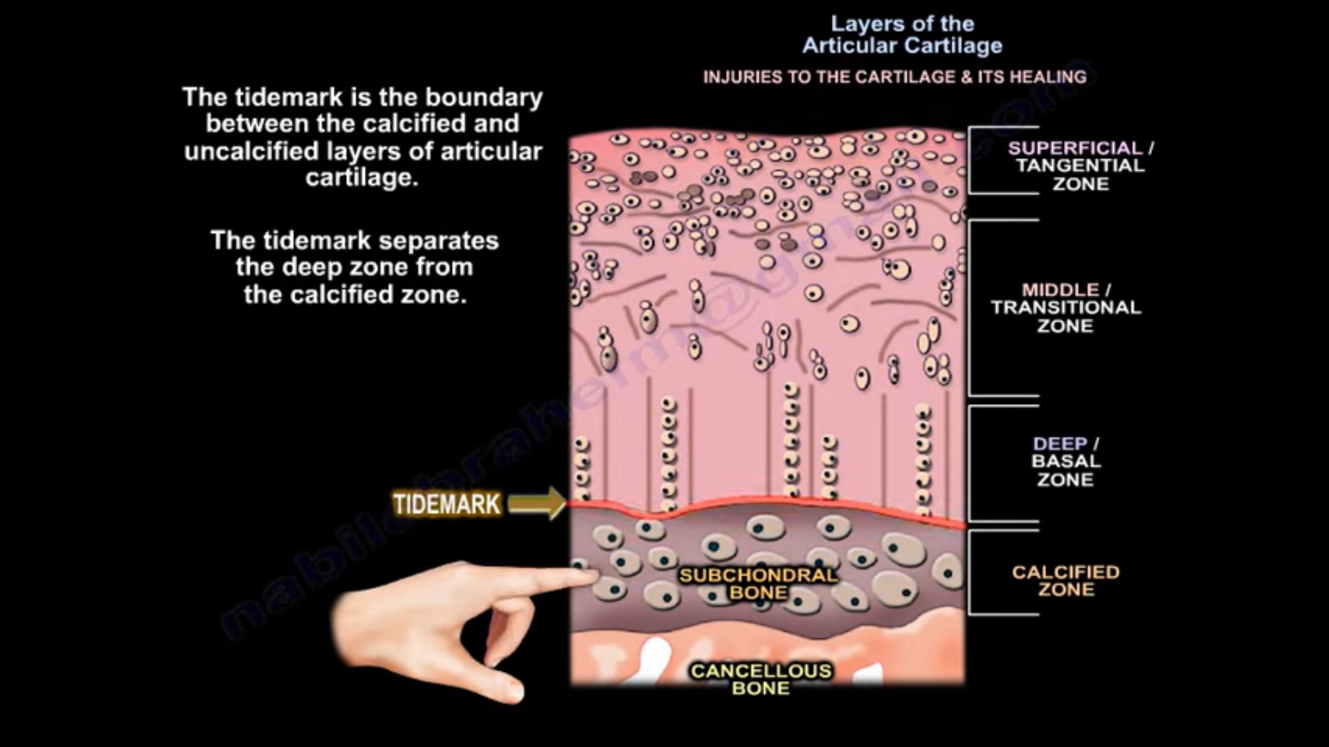
Articular cartilage itself is composed of four distinct layers:
-
Superficial (Tangential) Layer
-
Middle (Transitional) Layer
-
Deep (Radial) Layer
-
Calcified Zone
The Tidemark
-
The tidemark represents the boundary between:
-
Uncalcified cartilage (deep layer)
-
Calcified cartilage
-
-
It separates the deep zone from the calcified zone.
-
This junction is critical in determining the healing potential of cartilage injuries.
Collagen Composition of Cartilage
-
Normal articular cartilage is composed predominantly of Type Two collagen.
-
Fibrocartilage, which forms during repair after certain injuries, is composed mainly of Type One collagen.
-
Fibrocartilage is mechanically inferior to native articular cartilage.
Healing Response Based on Depth of Injury
1. Minimal Superficial Abrasion or Laceration
-
These injuries involve only the superficial layer of cartilage.
-
There is:
-
No hemorrhage
-
No inflammatory response
-
Minimal or no repair
-
-
Limited chondrocyte proliferation may occur near the injury site.
-
These lesions do not heal due to lack of blood supply.
2. Partial Thickness Cartilage Injury (Above the Tidemark)
-
Injuries involving up to fifty percent of cartilage thickness:
-
The remaining cartilage cannot adequately support mechanical stress.
-
Progressive degeneration occurs.
-
-
The damaged cartilage may transform into fibrocartilage, which is inferior in quality.
-
Chondrocytes may proliferate and synthesize matrix at the edges of the defect only.
-
There is no true repair of the defect.
3. Full Thickness Cartilage Injury Without Bone Involvement
-
Lesions that do not cross the tidemark:
-
Do not reach the subchondral bone
-
Do not access circulation
-
-
These injuries are unlikely to heal.
4. Osteochondral Injury (Crossing the Tidemark)
-
When the defect extends below the tidemark into the subchondral bone:
-
Blood supply is accessed
-
Mesenchymal stem cells are recruited
-
-
Healing occurs through the formation of fibrocartilage.
-
The repair tissue contains abundant Type One collagen.
-
Although healing occurs, the resulting cartilage is biomechanically inferior to native articular cartilage.
Key Characteristics of Deep Cartilage Lesions in Adults
-
Lesions crossing the tidemark:
-
Heal by fibrocartilage formation
-
Contain predominantly Type One collagen
-
-
The repair tissue lacks the durability and resilience of native articular cartilage.
Summary
-
Articular cartilage lesions that do not penetrate the subchondral bone do not heal due to the avascular nature of cartilage.
-
Partial thickness cartilage injuries result in limited cellular response without true repair.
-
Lesions that penetrate the subchondral bone can heal by recruiting mesenchymal stem cells.
-
Healing in such cases occurs through fibrocartilage formation, which is structurally and mechanically inferior to normal articular cartilage.
Leave a Reply