Courtesy: Matts Britberg, Sweden and ASPETAR, Qatar
Cartilage Injuries in Football: Epidemiology and Trauma Mechanisms
Introduction
Football is the most widely played sport in the world, with more than 300 million participants globally. As participation and athletic intensity continue to increase, the burden of musculoskeletal injuries—particularly articular cartilage injuries—has become increasingly significant.
Cartilage lesions are an important cause of:
- Pain
- Functional limitation
- Reduced athletic performance
- Early osteoarthritis
The knee, ankle, and hip are the most commonly affected joints in football players.
Epidemiology of Cartilage Injuries in Football
Incidence
Articular cartilage injuries are highly prevalent in athletes.
Reported findings include:
- Approximately 36% incidence of cartilage injuries in athletes
- Cartilage lesions identified in nearly 63% of arthroscopic procedures
- Full-thickness cartilage defects in approximately 17% of elite football players
Commonly Affected Joints
The joints most frequently involved are:
- Knee
- Ankle
- Hip
These joints are exposed to:
- Repetitive loading
- Rotational stress
- High-impact trauma
during football activity.
Risk Factors
Several factors increase the risk of cartilage injury in football players.
Player-Related Factors
- Higher body mass index (BMI)
- Elite-level participation
- Previous knee surgery
Surgical Risk Factors
Previous meniscectomy is particularly important because loss of meniscal tissue results in:
- Increased joint loading
- Reduced shock absorption
- Accelerated cartilage degeneration
Position-Specific Risk
Certain playing positions may experience:
- Higher repetitive loading
- Greater collision frequency
- Increased rotational stress
leading to a greater risk of cartilage damage.
Long-Term Consequences
Osteoarthritis Risk
Football players have a significantly increased risk of osteoarthritis.
Key observations include:
- Osteoarthritis risk up to 12 times higher than the general population
- Development of osteoarthritis approximately 4–5 years earlier
- Up to 32% of footballers eventually develop osteoarthritis
Functional Impact
Cartilage degeneration may lead to:
- Chronic pain
- Reduced athletic performance
- Disability
- Early retirement from sport
Mechanisms of Cartilage Injury
Cartilage injuries in football generally occur through two major mechanisms.
Chronic Repetitive Loading
Repeated loading over time may result in:
- Progressive cartilage degeneration
A dose-response relationship exists:
Moderate Loading
- May stimulate adaptive cartilage thickening
Excessive Loading
- Leads to cartilage breakdown and degeneration
Acute Traumatic Injury
Single high-impact events can produce:
- Focal cartilage defects
- Osteochondral fractures
- Cartilage fissures
- Matrix disruption
Common mechanisms include:
- Tackling injuries
- Twisting movements
- Pivoting
- Landing after jumping
Pathophysiology of Cartilage Damage
Excessive mechanical stress leads to biochemical cartilage degeneration.
Important pathological changes include:
- Decreased proteoglycan content
- Increased catabolic enzyme activity
- Chondrocyte apoptosis
These changes reduce the cartilage’s ability to:
- Resist load
- Maintain structural integrity
- Recover from repetitive stress
Types of Cartilage Lesions
Focal Cartilage Lesions
These are:
- Well-defined localized defects
Common causes include:
- Acute trauma
- Osteochondritis dissecans
- Osteonecrosis
Degenerative Cartilage Lesions
Degenerative lesions are:
- More diffuse
- Poorly defined
They are commonly associated with:
- ACL instability
- Meniscal injury
- Malalignment
Important Clinical Concept
Cartilage degeneration is not identical to osteoarthritis, but progressive degeneration may eventually lead to:
- Symptomatic osteoarthritis
Osteochondral Injuries
Definition
Osteochondral injuries involve damage to both:
- Articular cartilage
- Underlying subchondral bone
Clinical Features
These injuries may produce:
- Loose fragments
- Cartilage fissures
- Mechanical symptoms
Some lesions are subtle and may require:
- Arthroscopic probing for diagnosis
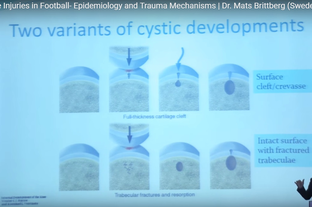
Subchondral Cyst Formation
Cartilage fissures may allow:
- Synovial fluid to enter subchondral bone
This can lead to:
- Formation of subchondral cysts
Patellar Dislocation and Cartilage Injury
Patellar dislocation is an important cause of osteochondral injury.
It is responsible for:
- Approximately 40–50% of femoral condyle cartilage injuries
These injuries frequently occur during:
- Twisting or pivoting movements
Role of Meniscal Injury
Meniscal damage contributes significantly to cartilage degeneration.
Loss of meniscal tissue results in:
- Increased contact pressure
- Reduced load distribution
- Accelerated cartilage wear
Biomechanics of Cartilage Injury
Rotational Forces
Shear and rotational stresses may produce:
- Cartilage matrix failure
These forces are common during:
- Pivoting
- Sudden direction changes
- Tackling
High-Impact Loading
When joint loading exceeds the cartilage tolerance threshold:
- Irreversible cartilage damage may occur
Injury Patterns in Football
Common football-related mechanisms include:
- Contact injuries
- Tackles
- Twisting injuries
- Landing injuries
Ankle Injuries
Most football-related ankle injuries involve:
- Inversion mechanisms
These injuries are strongly associated with later development of:
- Cartilage degeneration
- Osteochondral lesions
Prevention Strategies
Video Analysis
Video review helps identify:
- High-risk movements
- Mechanisms of injury
- Player-specific vulnerabilities
Modifiable Risk Factors
Several factors may be modified to reduce injury risk.
These include:
- Playing surface selection
- Footwear design
- Training modifications
- Load monitoring
Future Directions
Emerging concepts in injury prevention include:
- Biomarker screening
- Early cartilage damage detection
- Personalized injury prevention programs
These strategies may help identify:
- “At-risk” athletes
before irreversible cartilage damage develops.
Rehabilitation and Return to Sport
Successful recovery after cartilage injury requires:
- Structured rehabilitation
- Load management
- Progressive strengthening
- Sport-specific conditioning
Return-to-sport decisions should consider:
- Lesion severity
- Joint involved
- Surgical treatment
- Functional recovery
Key Clinical Pearls
- Cartilage injuries are extremely common in football players.
- The knee is the most frequently affected joint.
- Previous meniscectomy significantly increases cartilage degeneration risk.
- Both repetitive loading and acute trauma contribute to cartilage injury.
- Football players develop osteoarthritis earlier than the general population.
- Rotational shear forces are particularly damaging to cartilage.
- Prevention strategies should focus on biomechanics, training load, and early detection.
Final Take-Home Message
Cartilage injuries are a major source of long-term morbidity in football players and are strongly associated with early osteoarthritis and reduced athletic longevity.
Understanding:
- Epidemiology
- Biomechanics
- Injury mechanisms
- Risk factors
is essential for effective prevention, early diagnosis, and optimal management.
Early recognition and structured intervention can improve outcomes, preserve joint health, and prolong athletic careers.
Related Posts
- Neuromuscular Junction for FRCS Tr and Orth
Courtesy: Quen Tang, FRCS Orth, UK
 Cartilage Biology and Injuries
Courtesy: Dr Kunal Kalra Children’s Hospital of Michigan Pediatric Emergency Department, Michigan, USA www.chmpem.com
- Meniscus for FRCS Tr & Orth
Courtesy: Quen Tang, FRCS Orth
Leave a Reply