Courtesy: Christopher Gee, Golden Jubilee National Hospital and NHS Lanarkshire, UK
Early Onset Scoliosis
Courtesy: Muralidharan Venkatesan, Apollo Hospitals, Chennai
Overview of Distal Femur Fractures
Courtesy: Dr Yogesh Joshi, Consultant Knee Surgeon, Wrexham, UK
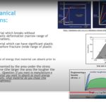
Biomaterials and Tribology for the FRCSOrth
Courtesy: Rishi Dhir, CEO, Lets Talk Dr, Consultant Orthopaedic Surgeon, Harlow, UK
Salter Harris Classification of Epiphyseal Injuries
Courtesy: Prof Nabil Ebraheim, University of Toledo, Ohio, USA GROWTH PLATE ANATOMY AND ITS FUNCTIONS INTRODUCTION The growth plate, also known as the physis, is the cartilaginous portion at the ends of long bones where longitudinal growth of the bone takes place. This region of bone is characterized by high metabolic activity and is under […]
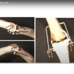
Hoffa fractures of the Distal Femur
COurtesy: Prof Nabil Ebraheim, University of Toledo, Ohio, USA
Update on Hip Arthroscopy

Courtesy: Oliver Marin-Pena, Madrid, Spain Hip Arthroscopy and Hip Preservation Surgery: Current Concepts and Practical Pearls Hip arthroscopy has become an increasingly important tool in the management of young adults with chronic hip and groin pain. Advances in imaging, surgical techniques, and understanding of hip biomechanics have expanded its indications. However, successful outcomes depend heavily […]
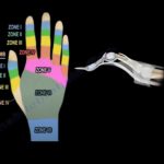
Applications of Wide awake Surgery in the Hand
Courtesy: The Master: Donald Lalonde ? Moderator: Sonu A. Jain ? Q&A: Peter Rhee ? Hand Therapist: Amanda Higgins Ashok Shyam, IORG, OrthoTV
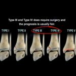
AcromioClavicular Joint Injury

Courtesy: Ali Noorani, Shoulder Surgeon, London, UK ACROMIOCLAVICULAR JOINT INJURIES Objectives ACJ Joint Anatomy & Stability Classification of ACJ Instability Management of Acute Injuries Management of Chronic Injuries Techniques for fixation Stability STATIC Every joint relies on Ligaments – acromioclavicular and coracoclavicular, bony Congruency and muscles around it in addition to a good functioning neurological […]
Boutonniere Deformity of Fingers
Courtesy: Prof Nabil Ebraheim, University of Toledo, Ohio, USA
Anatomy of Iliopsoas Muscle

Courtesy: Prof Nabil Ebraheim, University of Toledo, Ohio, USA ANATOMY OF ILIOPSOAS MUSCLE INTRODUCTION: IT INCLUDES 3 MUSCLES : PSOAS MAJOR,PSOAS MINOR(if present),ILIACUS. PSOAS MAJOR ORIGIN : It arises from the transverse processes and lateral aspect of vertebral bodies T12 – L5. COURSE :Runs downwards across the pelvic brim and then passes deep to the […]
Anatomy of The Gluteus Maximus Muscle
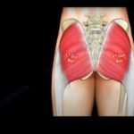
Courtesy: Prof Nabil Ebraheim, University of Toledo, Ohio, USA GLUTEUS MAXIMUS Gluteus maximus is the largest and heaviest muscle in the body. It is the most superficial of all gluteal muscles that are located at the posterior aspect of hip joint. ORIGIN: The gluteus maximus originates from The gluteal surface of ilium Lumbar fascia Sacrum […]
Congenital Hand Differences
Courtesy: NYU Langone Medical Centre, NYC Wee Lam Gill Smith Marybeth Ezaki Andrea Bauer
Basic science for Orthopaedic Boards
Courtesy: Amr Abdelgawad, Maimonaides Medical Centre, Brooklyn, NYC, USA
Anatomy Of The Tensor Fascia Lata Muscle
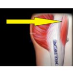
Courtesy: Prof Nabil Ebraheim, University of Toledo, Ohio, USA Tensor fascia lata muscle lies between tendon of gluteus maximus and tensor fascia lata at the middle of upper area of the thigh. Origin – anterior part of outer lip of iliac Crest Insertion – iliotibial tract at middle third of thigh (iliotibial band) Function: Flexion […]