Courtesy: Dr Cassandra A. Lee Ashok Shyam, OrthoTV
The Stem Cell Story

Courtesy: Dr Dominic Power, Consultant Hand Surgeon, Birmingham Hand Centre, United Kingdom Lecture delivered at International Bone Research Association Stem Cell Symposium, Sultan Qaboos University, Muscat, Oman, January 2014
Epidemiology & Classification of Spinal Cord Injury
Discussion on Epidemiology and Classification of Spinal Cord injury. Also assessment of hand function in tetraplegia Courtesy: Dominic Power, Consultant Hand Surgeon, Birmingham Hand Centre, UK
Anatomy of the Meniscus
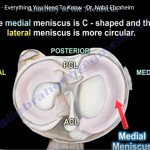
Courtesy: Prof Nabile Ebraheim, University of TOledo, Ohio, USA ANATOMY OF MENISCUS The meniscus is a cushion like structure made of cartilage which fits within the knee joint between the tibia and femur. There are two menisci inside the knee joint: 1. Medial meniscus 2. Lateral meniscus Medial meniscus is C shaped and the lateral […]
Kohler’s Disease
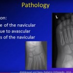
Courtesy: Dr Amr Abdelgawad, University of Texas, USA
Quick Rotational Profile
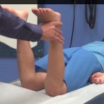
Courtesy: www.global-help.org Dr. Tom Jinguji, MD demonstrates the quick and easy exam — originally created by Dr. Lynn Staheli, MD.
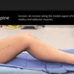
Tibial Spine Fractures in Children
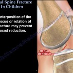
TIBIAL SPINE FRACTURES IN CHILDREN Fracture of tibial spine occur in skeletally immature patients It is similar to ACL injury in adults Mechanism : hyperextension injury of knee. Most common presentation is giving a history of fall from bicycle ,so an injured child who is brought to the emergency /OP with complaints of pain and […]
Distal #Clavicle Osteolysis
Courtesy: Prof Nabile Ebraheim, University of Toledo, Ohio, USA
Nerve Transfers in Partial Upper #Brachial Plexus Injuries
Courtesy: Dr Dominic Power FRCS, Birmingham Hand Centre, UK
How to avoid complications in Forearm Fractures in Children?
Courtesy: Atul Bhaskar, Ashok Shyam, IORG, OrthoTV
McMurray’s Test
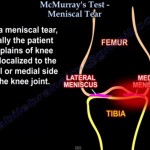
Courtesy:Prof Nabile Ebraheim, University of Toledo, Ohio, USA
Guest #Editor: May- August 2015- Cyril Mauffrey
Guest Editor: May 2015- August 2015 Dr Cyril Mauffrey MD, FACS, FRCS Director of Orthopaedic Trauma Denver Medical Centre, Denver, Colorado, USA • Dr Mauffrey is currently Associate Professor and Director of Orthopaedic Trauma at Denver Medical Centre. Currently the Chief Editor of The European Journal of Orthopaedic Surgery and Traumatology (Springer), his focus has […]
Saphenous Nerve Release
Saphenous Nerve Release Standard Edition (150401.150403) Courtesy: Authors: Susan E. Mackinnon, Andrew Yee Affiliation: Washington University School of Medicine Division of Plastic Reconstructive Surgery, Department of Surgery, Saint Louis, MO Peripheral Nerve Surgery: http://nervesurgery.wustl.edu Entrapment of the saphenous nerve is probably under-recognized and presents with numbness/pain in the infrapatellar region and the medial aspect of […]
Congenital Muscular Torticollis
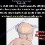
? CONGENITAL MUSCULAR TORTICOLLIS Usually caused by contracture of the sternocleidomastoid muscle Usually occurs in infants. It’s a common neck problem in childhood Cause is unknown – May be caused form pressure on the muscle or compartment syndrome of the muscle. Child holds the head towards the affected side with the chin rotated towards […]
The Sternocleidomastoid Muscle
Courtesy: Prof Nabile Ebraheim, University of Toledo, Ohio, USA STERNOCLEIDOMASTOID MUSCLE ANATOMY The sternocleidomastoid muscle (also known as sternomastoid) is one of the largest and most superficial cervical muscles located in the superficial layers on the side of the neck. ORIGIN The sternocleidomastoid muscle arises from the medial portion of the clavicle and the […]