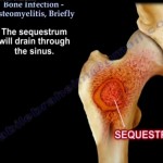
Courtesy: Prof Nabile Ebraheim, University of Toledo, Ohio, USA The video describes about Osteomyelitis and it’s management.It is an infection of bone in which bacteria enters the bone and they further attract leukocytes to the area which secretes enzymes in an attempt to destroy them.As a result blood flow to the area is reduced and […]
Bunionectomy and Hallux Valgus correction
Courtesy: Jeffrey Oster, DPM www.myfootshop.com
Ranawat Travelling Fellowship
Applications are invited for Ranawat Traveling Fellowship, which is co-funded by Rothman Institute, Dr. Chitranjan S. Ranawat, and others Eligibility: open to four (4) young orthopedic surgeons from throughout the world (including North America). The traveling Fellows will visit up to twelve (12) host sites in North America within a period of approximately four (4) […]
Syndactyly Classification
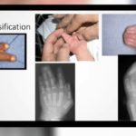
Syndactyly is the most common congenital hand anomaly Acrosyndactyly: fusion of distal parts of the digit with proximal fenestration Isolated syndactyly has an autosomal dominant inheritance with variable penetrance Frequency of syndactyly: thumb-index (5%), index-middle (15%), middle-ring (50%), ring-little (30%) Type Characteristics Simple […]
Fellowship in Paediatric Orthopaedics
OrthoKids Clinic, Ahmedabad invites applications for Clinical Fellow position in Pediatric Orthopedics. OrthoKids Clinic is an established Pediatric Orthopedic Center for last nine years. It is a private set up where all kinds of Pediatric Orthopedic elements are treated by Dr. Maulin Shah. There is a good turnover of patients providing an opportunity to observe […]
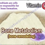
Vitamins D, E and K and Orthopaedics
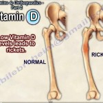
Courtesy:Prof Nabile Ebaheim, University of Toledo, Ohio, USA
Percutaneous Achilles Tendon Repair
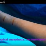
Courtesy: Gautam Salunkhe,Nirmala Hospital, hadapsar, Pune and Ashok Shyam, MOA, IORG and OrthoTV
Oswestry Shoulder and Elbow Course
OSWESTRY SHOULDER AND ELBOW COURSE Dates: July 1, 2015 – July 2, 2015 Location: Robert Jones & Agnes Hunt Orthopaedic Hospital NHS Foundation Trust, UK Website: http://www.orthopaedic-institute.org
European Bone and Joint Infection Society 39th Annual Meeting 2020
European Bone and Joint Infection Society Meeting Venue: GR – Ljubljana Exhibition & Convention Center Dunajska Cesta 18 Ljubljana, Slovenia, Europe Dates: 10-12 September 2020 Website: https://ebjis2020.org/ Last Date for Abstract Submission: 10 April 2020
AO Trauma Osteotomy Course
AO Trauma Osteotomy Course Venue: Radisson Blu Hotel, NewDelhi Dates: July 3, 4, 2015 Brochure
Vitamin A, B and C in #Orthopaedics
Courtesy: Prof Nabile Ebraheim, Unviersity of Toledo, Ohio, USA
Linburg Comstock Anomaly and Median nerve symptoms
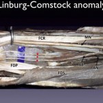
Courtesy: Dr Dominic Power, Birmingham Hand Centre, UK
THA in Pelvic Discontinuity
Courtesy: Dr. Anil Oommen, Dr Ashok Shyam, IORG, OrthoTV
Four Corner Arthrodesis for SNAC wrist using a variable angle locking plate
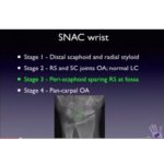
Courtesy: Dr Dominic Power, FRCS, Consultant Hand Surgeon, Birmingham Hand Centre, UK
Modified Osteocutaneous Foucher Flap
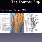
Courtesy: Dr Dominic Power, FRCS, Birmingham Hand Centre, UK