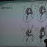
Courtesy: Dr DC Sundaresh, Chairman, Orthopaedics, Sri Sathya Sai Institute of Higher Medical Science, AP
Cubital Tunnel Syndrome
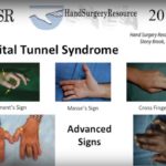
Courtesy: Larry Hurst MD, Stony Brook University Hospital, NY, USA and Hand Surgery Resources
Innovations in Total Hip Replacement
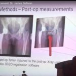
Courtesy: James Sullivan, Head of Athroplasty. The Australian School of Advanced Medicine, Macquaire University, Australia and Sri Sathya Sai Institute of Higher Medical Science, AP
Principles of Buttress Plating
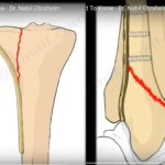
Courtesy: Prof Nabil Ebraheim, University of Toledo, Ohio, USA
Bridge Plating Concepts in Fracture Healing
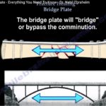
Courtesy: Prof Nabil Ebraheim, University of Toledo, Ohio, USA
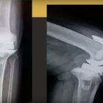
Atypical femur fractures – where are we now?
Courtesy: Saqib Rehman, OrthoClips www.orthoclips.com
Ankle Fractures – Avoiding the Landmines
Courtesy: Saqib Rehman, Temple University, Philadelphia, USA
Anatomy of Flexor Digitorum Superficialis
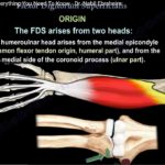
Courtesy: Prof Nabil Ebraheim, University of Toledo, Ohio, USA ANATOMY OF FLEXOR DIGITORUM SUPERFICIALIS Flexor digitorum superficialis is a superficial digit flexor muscle that covers radius,ulna and flexor digitorum profundus ORIGIN a)Humeroulnar head -from medial epicondyle and from medial side of coronoid process of ulna b)Radial head from oblique upper third of anterior border of […]
Anatomy of Obturator Artery

Courtesy: Prof Nabil Ebraheim, University of Toledo, Ohio, USA Overview The obturator artery is a key pelvic vessel supplying: The medial thigh Structures around the hip joint Clinical Importance Understanding its anatomy is essential in: Pelvic trauma Acetabular fracture surgery Total hip arthroplasty Anatomical variations can lead to significant hemorrhage if injured Origin of the […]
Proximal humerus fractures – Fix or replace?
Courtesy: Saqib Rehman, Temple University, Philadelphia, USA
Difference between Orthopaedics and Orthopedics
What is the Difference between Orthopaedics and Orthopedics? The words Orthopaedics and Orthopedics are used interchangeably, they imply the same, the speciality of Medicine dealing with the treatment of Bone and Joint Disorders “Orthopaedics” is used in a British context and “Orthopedics” from an American context Interestingly, the American Academy of Orthopaedic Surgeons use the […]
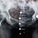
Robotic Assisted Spine Surgery
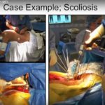
Courtesy: Isador Lieberman MD, Texas Back Institute
Anatomy of Quadratus Femoris
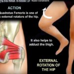
Courtesy: Prof Nabil Ebraheim, University of Toledo, Ohio, USA ANATOMY OF QUADRATUS FEMORIS MUSCLE ORIGIN From the lateral margin of ischial tuberosity INSERTION Into the quadrate tubercle on the intertrochanteric crest between the greater and lesser trochanter ACTION External rotation of the hip and also helps in adduction of thigh NERVE SUPPLY The nerve from […]
Arthroscopic Eden-Hybinette procedure with autograft
Courtesy: Ettore Taverna and Emmanuel Brilakis, Greece and Athens Shoulder Course
Measured Resection Vs Gap Balancing technique in TKR
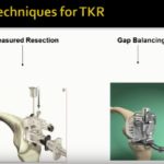
Courtesy: Paul D Maitino, Oklahoma, USA