Courtesy: Dr Taral Nagda, Paediatric Orthopaedic Surgeon, Saifee Hospital, Mumbai
Osteoclasts for the FRCSOrth
Courtesy: Quen Tang, FRCSOrth, UK
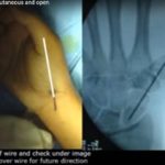
Percutaneous and Open techniques for Bennet fracture
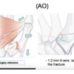
Courtesy: Binu Prathap Thomas, Professor of Hand Surgery, Christian Medical College, Vellore, India
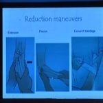
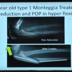
The Enigmatic Elbow and TRASH Lesions
Courtesy: Sandeep Patwardhan, Ashok Shyam, Sancheti Institute, Pune and IORG, OrthoTV
Clinical Examination of the Shoulder Joint
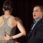
Courtesy: Brian Feeley MD, UCSF Orthopedics of San Francisco, CA
Current Trends in Anatomical Shoulder Replacement
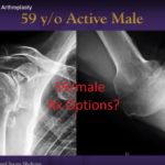
Courtesy: Jason Hsu and Ahsan MD, University of Washington, Seattle, USA
Radiographic Assessment of Bone Lesions
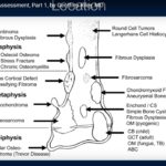
Part 1 of Lecture Part 2 of Lecture Courtesy: Geoffrey Riley MD, Associate Professor and Consultant Radiologist, Stanford University, USA
Anatomy and Contents of the Anatomical Snuff Box
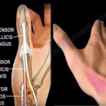
Courtesy: Prof Nabil Ebraheim, University of Toledo, Ohio, USA The anatomical snuffbox is a small triangular depression located on the dorsoradial aspect of the wrist.People used this space to place and sniff the powdered tobacco or “snuff”, hence the name.The base of this triangular space is proximal with the apex pointing towards the thumb.The anatomical […]
Anatomy and Contents of the Cubital Fossa
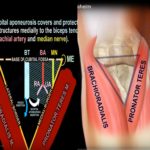
Courtesy: Prof Nabil Ebraheim, University of Toledo, Ohio, USA The cubital fossa is a triangular depression located in front of the anterior elbow.The medial border is formed by the pronator teres which arises from the medial epicondyle of the humerus.The lateral border is formed by the brachioradialis muscle which arises from the lateral supracondylar ridge […]
All Inside ACL Reconstruction
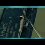
Courtesy: Dr Deepak Joshi, Safdarjung Hospital, NewDelhi
Radiographic Imaging of the Hip Joint
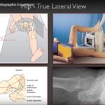
Courtesy: Christopher Beaulieu, Professor of Radiology, Stanford University, USA
Congenital Talipes Equinovarus
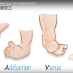
Courtesy: Sameer Qureshi, Consultant Orthopaedic Surgeon, The Young Orthopod Channel
Radiography of Arthritis
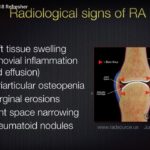
Courtesy: Christopher Beaulieu, Professor of Radiology, Stanford University, USA
Joint Dislocations for the FRCS Orth
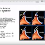
Courtesy: FRCS Orth Preparatory course
Paediatric Ortho for the FRCS Tr and Orth Exit exam
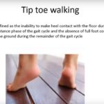
Courtesy: Shanaka serevirathna FRCS Tr and Orth, FRCS Orth Preparatory Course