• Skip to main content
  • Skip to secondary menu
  • Skip to primary sidebar
OrthopaedicPrinciples.com

OrthopaedicPrinciples.com

Integrating Principles and Evidence

Integrating Principles and Evidence

  • Home
  • Editorial Board
  • Our Books
    • Evidence Based Orthopaedic Principles
  • Courses
  • Exams
  • Reviews
  • Live Program
  • Contact

Anatomy of the Extensor Indicis Muscle

Courtesy: Prof Nabil Ebraheim, University of Toledo, Ohio, USA

Anatomy of the Extensor Indicis Muscle

Overview

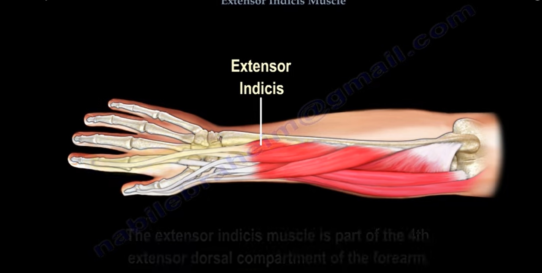
  • The extensor indicis muscle is part of the fourth dorsal extensor compartment of the forearm.

  • It lies deep to the extensor digitorum communis muscle.

  • It has the most distal muscle belly within the fourth dorsal compartment.

  • This distal muscle belly is clinically important, as it helps in identification of the extensor indicis muscle during tendon lacerations involving multiple extensor tendons.

  • The tendon of the extensor indicis lies ulnar to the extensor digitorum communis tendon to the index finger.


Origin

  • Posterior surface of the ulna

  • Interosseous membrane


Insertion

  • Extensor expansion of the index finger


Innervation

  • Posterior interosseous nerve


Function

  • Extension of the index finger

  • Assists in extension of the wrist


Examination of the Extensor Indicis Muscle

  • The muscle is examined by:

    • Asking the patient to make a fist

    • Then asking the patient to extend the index finger independently


Tendons Responsible for Index Finger Extension

  • Two tendons contribute to extension of the index finger:

    • Extensor indicis muscle

    • Extensor digitorum communis tendon to the index finger


Indications for Tendon Transfer Using Extensor Indicis

  • Rupture of the extensor pollicis longus tendon

    • Extensor pollicis longus tendon may rupture following a nondisplaced fracture of the distal radius

    • This results in inability to extend the thumb

    • The extensor indicis tendon can be transferred to restore thumb extension

  • Median nerve injury

    • Extensor indicis opponensplasty may be performed to restore thumb opposition


Principles of Performing Extensor Indicis Tendon Transfer

  • During tendon transfer:

    • The extensor indicis tendon should be sectioned proximal to the sagittal hood

    • This preserves independent extension of the index finger through the extensor digitorum communis


Contraindications for Extensor Indicis Transfer

  • In cases of rheumatoid arthritis:

    • The extensor digitorum communis tendon to the index finger may already be ruptured

    • In such situations, transfer of the extensor indicis tendon is contraindicated, as it would eliminate index finger extension


Key Take-Home Points

  • Extensor indicis provides independent extension of the index finger

  • It is a reliable donor tendon for reconstruction

  • Proper identification and preservation of index finger extension are essential during tendon transfer

Post Views: 2,204

Related Posts

  • Anatomy of the #Extensor Indicis Muscle

    Courtesy: Prof Nabile Ebraheim, University of Toledo, Ohio, USA

  • Anatomy Of The Extensor Digitorum Longus Muscle

    Courtesy: Prof Nabil Ebraheim, University of Toledo, Ohio, USA

  • Anatomy of Plantaris Muscle

    Courtesy: Prof Nabil Ebraheim, University of Toledo, Ohio, USA

Reader Interactions

Leave a Reply

Your email address will not be published. Required fields are marked *

Primary Sidebar

Follow Us

instagram slideshare

Categories

  • -Applied Anatomy
  • -Approaches
  • -Basic Sciences
  • -Cartilage & Meniscus
  • -Classifications
  • -Examination
  • -Foot and Ankle
  • -Foot and Ankle Trauma
  • -FRCS(Tr and Orth) tutorials
  • -Gait
  • -Hand and Wrist
  • -Hand and Wrist Trauma
  • -Hand Infections
  • -Hip and Knee
  • -Hip Preservation
  • -Infections
  • -Joint Reconstruction
  • -Knee Arthroplasty
  • -Knee Preservation
  • -Metabolic Disorders
  • -Oncology
  • -OrthoBiologics
  • -OrthoPlastic
  • -Paediatric Orthopaedics
  • -Paediatric Trauma
  • -Patellofemoral Joint
  • -Pelvis
  • -Peripheral Nerves
  • -Principles
  • -Principles of Surgery
  • -Radiology
  • -Rheumatology
  • -Shoulder and Elbow
  • -Shoulder and Elbow Arthroplasty
  • -Spine Deformity
  • -Spine Oncology
  • -Spine Trauma
  • -Spine, Pelvis & Neurology
  • -Sports Ankle and Foot
  • -Sports Elbow
  • -Sports Knee
  • -Sports Medicine
  • -Sports Medicine Hip
  • -Sports Shoulder
  • -Sports Wrist
  • -Statistics
  • -Technical Tip
  • -Technology in Orth
  • -Trauma
  • -Trauma (Upper Limb)
  • -Trauma Life Support
  • -Trauma Reconstruction
  • Book Shelf
  • Book Shelf Medical
  • Careers
  • Case Studies and Free Papers
  • DNB Ortho
  • Evidence Based Orthopaedic Principles
  • Evidence Based Orthopaedics
  • Exam Corner
  • Fellowships
  • Guest Editor
  • Guest Reviews
  • Image Quiz
  • Instructional Course Lectures
  • Journal Club
  • MCQs
  • Meetings and Courses
  • Multimedia
  • News and Blog
  • Plaster Techniques
  • Podcasts
  • Public Health
  • Rehabilitation
  • Research
  • Shorts and Reels
Copyright@orthopaedicprinciples.com. All right rerserved.