Courtesy Dr Sachin Tapasvi, Dr Ashok Shyam, Ortho TV
ACL Reconstruction: Surgical Planning, Technique and Key Principles
Introduction
Anterior Cruciate Ligament (ACL) reconstruction is one of the most commonly performed procedures in sports orthopaedics. Successful outcomes depend not only on surgical skill, but also on appropriate patient selection, graft choice, accurate tunnel placement, biological preservation, and secure fixation.
Modern ACL surgery has evolved considerably with improved understanding of ACL anatomy and biomechanics, leading to more anatomical and individualized reconstruction techniques.
Patient Selection for ACL Reconstruction
Clinical Diagnosis First
ACL injury is primarily a clinical diagnosis. The decision to proceed with surgery depends on several factors, including:
- Symptomatic instability
- Functional limitations
- Failure of conservative treatment
- Activity demands of the patient
Patients with recurrent instability during sports or daily activities are more likely to benefit from reconstruction.
Preoperative Evaluation and Planning
Patient-Specific Factors
Preoperative planning must be individualized. Important considerations include:
Activity Level
- Sedentary individual versus athlete
- Participation in pivoting sports versus non-pivoting activities
Occupational Requirements
Certain occupations may require:
- Frequent kneeling
- Climbing
- Heavy manual activity
Cultural and Religious Needs
Activities involving kneeling or squatting may influence graft selection and rehabilitation goals.
Cosmetic Concerns
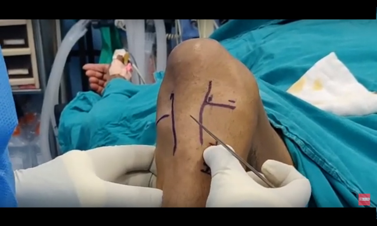
Incision placement and donor-site appearance may be important, especially in young patients.
Patient Expectations
Discussion should include:
- Return to sports
- Timing of return
- Desired activity level
- Daily functional goals
Previous ACL Surgery
Importance of Surgical History
If the patient previously underwent ACL reconstruction on the opposite knee, important questions include:
- Which graft was used?
- Was the patient satisfied with the outcome?
If prior results were satisfactory, using the same graft type may be advantageous.
Assessment of Associated Injuries
ACL tears are frequently associated with additional injuries that must be identified before surgery.
Common Associated Injuries
- Meniscal tears
- Collateral ligament injuries
- Posterolateral corner injuries
These may require:
- Combined procedures
- Staged reconstruction
Failure to address associated pathology may compromise surgical outcomes.
Graft Selection Strategy
ACL Footprint-Based Graft Selection
The size of the native ACL footprint influences graft choice.
Small Footprint (<16 mm)
Preferred grafts:
- Hamstring graft
- Five- or six-strand hamstring constructs
Large Footprint (>16 mm)
Preferred grafts:
- Bone–Patellar Tendon–Bone (BTB) graft
- Quadriceps tendon graft
Importance of Graft Size
Minimum Recommended Diameter
A graft diameter of at least 8–8.5 mm is generally recommended.
Smaller grafts are associated with:
- Higher re-rupture rates
- Increased graft failure risk
Adequate graft diameter is therefore critical for long-term success.
Graft Healing Considerations
Different graft types incorporate and mature at different rates.
This influences:
- Rehabilitation progression
- Return-to-sport timelines
Biological healing should always be considered when designing postoperative rehabilitation protocols.
Operating Room Setup and Surgical Principles
Equipment
ACL reconstruction is highly dependent on arthroscopic technology.
Essential requirements include:
- High-quality arthroscopy camera systems
- Reliable instrumentation
- Accurate drilling systems
Improved equipment contributes to greater surgical precision.
Patient Positioning
Positioning Techniques
Common methods include:
- Side post
- Leg holder systems
Proper positioning allows:
- Full knee manipulation
- Hyperflexion during femoral tunnel drilling
- Improved arthroscopic access
Sterility and Surgical Preparation
Preparation Sequence
Sequential preparation should proceed from:
- Foot
- Knee
- Entire limb
Modern sterility principles include:
- Impervious drapes
- Plasma sterilization
- Autoclave systems
Older toxic sterilization agents such as formalin chambers and glutaraldehyde should be avoided.
Arthroscopic Portal Placement
Three-Portal Technique
A three-portal technique is commonly recommended.
1. Anterolateral Portal
- Primary viewing portal
- Positioned high
2. Anteromedial Portal
- Viewing and working portal
- Positioned high and close to the medial patellar tendon border
3. Accessory Anteromedial Portal
- Main working portal
- Positioned low and medially
Principles of Portal Placement
Proper portal positioning improves:
- Instrument maneuverability
- Femoral tunnel orientation
- Tunnel length
- Arthroscopic visualization
A high anteromedial portal reduces crowding, while a low accessory portal improves access to the femoral footprint.
Femoral Tunnel Considerations
Influence of Portal Position
Portal placement affects femoral tunnel geometry.
More Lateral Anteromedial Portal
- Produces longer tunnels
More Medial Portal
- Creates shorter and more oblique tunnels
Ideal Femoral Tunnel
Desired Characteristics
An ideal femoral tunnel should:
- Measure approximately 35–40 mm in length
- Be anatomically positioned
- Preserve surrounding structures
Tunnel drilling is commonly performed with the knee hyperflexed beyond 120°.
Importance of Femoral Tunnel Position
Femoral tunnel malposition is one of the leading causes of ACL reconstruction failure, accounting for more than 40% of failed cases in some series.
Accurate tunnel positioning is therefore one of the most important technical aspects of surgery.
Visualization Principles
Viewing the femoral footprint from the anteromedial portal provides superior visualization and improves understanding of native ACL anatomy.
Switching viewing portals during surgery may further improve anatomical orientation.
Step-by-Step Surgical Technique
1. Diagnostic Arthroscopy
Initial arthroscopy is used to:
- Confirm ACL tear
- Evaluate remnant tissue
- Identify associated injuries
Useful remnant tissue should be preserved whenever possible.
2. Femoral Side Preparation
The femoral footprint is identified using:
- ACL stump remnants
- Anatomical landmarks
A radiofrequency probe may be used for marking the footprint. Tunnel size is matched to graft diameter.
3. Femoral Tunnel Creation
Steps include:
- Insert guide pin
- Drill tunnel in hyperflexion
- Overdrill according to graft size
- Remove tunnel debris
- Measure tunnel length
- Pass shuttle suture
4. Tibial Tunnel Creation
Tunnel Placement
The tibial tunnel is generally positioned:
- Slightly medial to center
- Approximately at the medial two-fifths of the footprint
Technical Considerations
- Use serial drilling
- Avoid cartilage injury
Graft Passage
The graft is passed through the tibial and femoral tunnels while ensuring:
- Proper seating
- Correct orientation
- Smooth passage without twisting
Graft Fixation
Graft Cycling
The knee is cycled approximately 20 times between:
- 0° and 90° flexion
Purpose:
- Remove collagen creep
- Precondition the graft
Femoral Fixation Methods
Fixation method depends on graft type.
Soft Tissue Grafts
Options include:
- Interference screws
- Suspensory fixation devices
Bone–Patellar Tendon–Bone Grafts
Metal interference screws are often preferred for bone plug fixation.
Tibial Fixation
Knee Position During Fixation
Fixation is generally performed with the knee at:
- Approximately 20° flexion
In patients with significant hyperextension:
- Fixation in full extension may be preferred
A posterior drawer force is applied before final fixation.
Screw Size Selection
General guidelines include:
- If the screwdriver fits snugly: use screw size +1 mm
- In soft bone: use +2 mm screw
- In very tight tunnels: use same-sized screw
Proper screw sizing improves fixation stability.
Key Surgical Principles
Important principles include:
- Minimum graft diameter of at least 8–8.5 mm
- Coverage of at least 80% of the native ACL footprint
- Use of a three-portal technique
- Visualization of the femoral footprint through the anteromedial portal
- Anatomical tunnel placement
- Preference for transportal or outside-in drilling techniques
Post-Fixation Rehabilitation
Following stable fixation of an isolated ACL reconstruction:
- Accelerated rehabilitation protocols may be initiated
Rehabilitation should still respect graft biology and healing timelines.
Key Takeaways
Successful ACL reconstruction requires a patient-specific approach.
Critical factors include:
- Appropriate graft choice
- Accurate tunnel placement
- Adequate graft size
- Secure fixation
- Recognition and treatment of associated injuries
The most common technical cause of failure remains femoral tunnel malposition. Careful surgical planning and precise execution are therefore essential for optimal outcomes.


Leave a Reply