
Courtesy: Gustavo Gomez Rodriguez, Buenos Aires, Argentina
Wrist Arthroscopy: Step by Step
Overview
- Wrist arthroscopy is a minimally invasive technique that allows direct visualization of the structures within the wrist through small portals.
- Compared with conventional open approaches, arthroscopy can reduce soft-tissue disruption and may facilitate recovery in appropriately selected patients.
- The technique can be used both for diagnosis and treatment, including assessment of cartilage, ligament injuries, fractures, and instability.
- A structured learning process is important because wrist arthroscopy requires familiarity with three-dimensional anatomy, portal placement, triangulation, and instrument handling.
Wrist Arthroscopic Portals
- The wrist contains six dorsal extensor compartments, and several commonly used arthroscopic portals are named according to their relationship to these compartments.
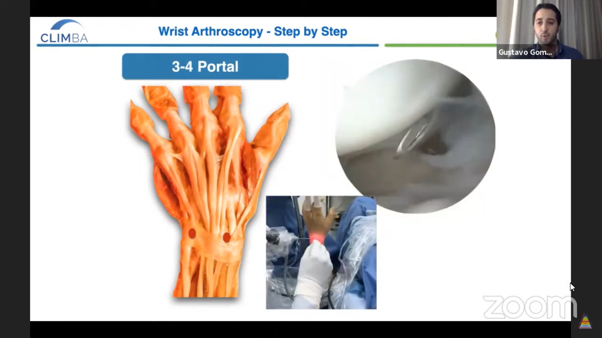
- The 3–4 portal is located between the third and fourth extensor compartments and is one of the principal portals used for radiocarpal arthroscopy.
- The 6R portal is positioned on the radial side of the extensor carpi ulnaris tendon and provides another important access point to the radiocarpal joint.
- Additional portals include the radial and ulnar midcarpal portals.
- Less commonly used portals may be selected according to the pathology being evaluated or treated.
Basic Arthroscopic Examination
- Portal placement is generally performed using a needle to identify the joint before making a small skin incision.
- A blunt instrument can then be used to safely enter the capsule before introducing the arthroscope.
- The radiocarpal joint provides visualization of important structures, including:
- The lunate.
- The proximal pole of the scaphoid.
- The scaphoid and lunate facets of the distal radius.
- Examination can subsequently be extended to the midcarpal joint.
- With appropriate experience, a systematic examination of the radiocarpal and midcarpal joints can be performed efficiently.
- Direct visualization can provide information that may not be fully apparent on radiographs or fluoroscopy.
Developing Arthroscopic Skills
A progressive approach to training can help surgeons develop the necessary skills.
Early-Level Procedures
- Arthroscopic treatment of selected dorsal wrist ganglia.
- Arthroscopic assistance during relatively straightforward distal radius fractures.
- Arthroscopic assessment of suspected ligament injuries.
Intermediate-Level Procedures
- Arthroscopic assistance in more complex distal radius fractures.
- Treatment of selected triangular fibrocartilage complex injuries.
- Arthroscopic assistance during carpal fracture fixation.
Advanced Procedures
- Complex distal radius fractures.
- Complex ligament reconstruction.
- Selected distal radius malunions.
- Arthroscopic procedures involving basal thumb arthritis.
Cadaveric training and repeated practice can help develop triangulation, suturing, portal orientation, and instrument-control skills before undertaking complex clinical procedures.
Arthroscopic Treatment of Dorsal Wrist Ganglia
- Arthroscopic excision is an option for appropriately selected dorsal wrist ganglia.
- Potential advantages include smaller incisions and reduced soft-tissue disruption.
- The arthroscope and shaver can be introduced through appropriately selected portals.
- The origin or stalk of the ganglion is identified arthroscopically.
- Resection of the associated capsular tissue can be performed around the stalk.
- The procedure is continued until the ganglion’s communication with the joint has been adequately addressed.
- Careful identification of the surrounding extensor tendons is important during the procedure.
Arthroscopic Assistance in Distal Radius Fractures
- Arthroscopy can be particularly useful when a distal radius fracture involves the articular surface.
- Important goals of fracture treatment include restoration of:
- Articular congruity.
- Radial alignment.
- Volar tilt.
- Ulnar variance.
- Arthroscopy allows direct inspection of the articular surface and can help identify residual steps or gaps following fracture reduction.
- A probe may be used to manipulate or assess articular fragments.
- Temporary Kirschner wires can maintain reduction before definitive fixation.
- In selected impacted fractures, fragments can be elevated from beneath the articular surface through a cortical window before fixation.
- Arthroscopy can also reveal associated cartilage injury resulting from the original trauma.
Assessment of Scapholunate Ligament Injuries
- Wrist arthroscopy provides a direct method of assessing the integrity of the scapholunate ligament.
- Arthroscopic staging systems can help characterize the severity of scapholunate injury.
- In less severe injuries, a probe may enter the interval between the scaphoid and lunate without substantial instability.
- As instability progresses, the probe may pass more freely through the scapholunate interval.
- In advanced injuries, the arthroscope itself may be able to pass through the scapholunate interval between the radiocarpal and midcarpal compartments.
- Arthroscopy therefore provides useful information about the degree of ligament disruption and carpal instability.
Assessment of the Triangular Fibrocartilage Complex
- The triangular fibrocartilage complex is an important stabilizer of the distal radioulnar joint.
- Its peripheral attachments include superficial and deep components.
- The deep radioulnar ligament attachments at the ulnar fovea are particularly important for distal radioulnar joint stability.
- Arthroscopic examination can be used to assess the integrity of these attachments.
- The hook test can help evaluate the stability of the triangular fibrocartilage complex and its foveal attachment.
- The trampoline test assesses the tension and stability of the triangular fibrocartilage complex.
- These tests can help distinguish between stable and unstable peripheral triangular fibrocartilage complex injuries.
Arthroscopic-Assisted Triangular Fibrocartilage Complex Repair
- When the triangular fibrocartilage complex tissue is of adequate quality, repair may be considered for selected peripheral or foveal tears.
- Foveal repair aims to restore the attachment of the deep triangular fibrocartilage complex to the ulna.
- One described approach uses transosseous tunnels through the ulnar foveal region.
- Sutures can be passed through the triangular fibrocartilage complex and secured to restore the tissue to its anatomical attachment.
- A successful repair should improve the stability of the distal radioulnar joint.
- Arthroscopy allows direct assessment of the repair and can be used to reassess the triangular fibrocartilage complex after fixation.
When Reconstruction Is Required
- Primary repair is dependent on having adequate-quality tissue.
- Chronic injuries, severely damaged tissue, or irreparable triangular fibrocartilage complex tears may require reconstruction rather than simple repair.
- Tendon graft reconstruction is one possible approach in appropriately selected patients.
- The specific reconstruction technique should be selected according to the pattern of instability, tissue quality, associated deformity, and surgeon experience.
Arthroscopic Assistance in Scaphoid Fracture Fixation
- Scaphoid fractures are among the most common carpal fractures requiring surgical treatment.
- Arthroscopy can assist with reduction and provide direct visualization of the articular surfaces.
- It can also help confirm appropriate positioning of fixation hardware.
- During screw fixation, arthroscopy may allow visualization of the screw trajectory and help identify potential penetration of the cartilage or joint surface.
- This can complement fluoroscopic imaging during fracture fixation.
Complex Distal Radius Fractures
- Arthroscopy can be used as an adjunct during fixation of complex intra-articular distal radius fractures.
- The objective is to restore the articular surface while maintaining the overall alignment of the distal radius.
- A systematic reduction strategy may involve addressing fragments sequentially rather than attempting to reduce all fragments simultaneously.
- Arthroscopic visualization helps identify:
- Articular steps.
- Residual gaps.
- Depressed fragments.
- Cartilage injuries.
- A temporary Kirschner wire can be used as a joystick to manipulate difficult fragments.
- Once satisfactory reduction has been achieved, temporary fixation can be replaced or supplemented with definitive locking fixation.
- The final arthroscopic assessment can confirm restoration of articular congruity.
Scapholunate Instability and Ligament Reconstruction
- Chronic scapholunate instability can produce characteristic changes in carpal alignment.
- Imaging may demonstrate:
- Scaphoid flexion.
- An increased scapholunate angle.
- Dorsal intercalated segment instability alignment.
- Abnormal positioning of the scaphoid relative to the distal radius.
- Magnetic resonance imaging can demonstrate ligament injury and associated soft-tissue abnormalities.
- Computed tomography can provide detailed information regarding carpal bone position and rotation.
- Dynamic fluoroscopic examination may also be useful for assessing carpal instability.
- Arthroscopy helps determine whether the carpal alignment can still be corrected before proceeding with reconstruction.
- In selected cases, minimally invasive ligament reconstruction can use suture tape or tendon graft techniques.
- Three-dimensional guides are being explored to improve the accuracy of bone tunnel placement in reconstructive procedures.
- Arthroscopic assessment after reconstruction can help confirm restoration of the relationship between the scaphoid and lunate.
Arthroscopic Management of Distal Radius Malunion
- Even relatively small articular incongruities can be clinically significant in some patients.
- Arthroscopy can help visualize an articular step and guide correction during selected corrective osteotomy procedures.
- An arthroscopic-assisted approach may allow the surgeon to directly assess the articular surface while mobilizing the malunited fragment.
- The fragment can be elevated and repositioned to restore a more congruent joint surface.
- Temporary wires and fragment-specific fixation can be used to maintain the corrected position.
- Final fixation may be completed using an appropriate locking plate.
- Arthroscopy provides an opportunity to assess residual articular incongruity and cartilage damage.
Basal Thumb Arthritis
- Arthroscopy also has a role in selected cases of thumb carpometacarpal joint arthritis.
- It may be considered in appropriately selected patients with earlier-stage disease and suitable joint pathology.
- Commonly described arthroscopic portals allow triangulation of instruments while minimizing the risk to nearby sensory nerve branches.
- A systematic inspection of the joint can be performed by examining different regions of the articular surface.
- Synovial tissue can be debrided when indicated.
- Osteophytes and loose bodies may be removed arthroscopically.
- The condition of the cartilage on the trapezium and base of the first metacarpal can be assessed directly.
- In selected patients, arthroscopic trapeziectomy can be performed.
- A burr may be used to remove the trapezium progressively, while fluoroscopy can help assess the extent of bone removal.
- The extent of resection can be tailored to the clinical and surgical requirements.
Arthroscopy for Distal Radioulnar Joint Instability
- In patients with distal radioulnar joint instability, assessment of the triangular fibrocartilage complex is particularly important.
- Foveal detachment of the triangular fibrocartilage complex may contribute significantly to instability.
- If the tissue remains sufficiently healthy, an anatomical repair may restore stability.
- When the tissue is irreparable or of poor quality, ligament reconstruction using a graft may be necessary.
- Reconstruction techniques may recreate the stabilizing attachments around the distal radioulnar joint.
- Treatment should be individualized according to the cause and chronicity of instability, tissue quality, bony anatomy, and associated injuries.
Arthroscopy in Acute Scapholunate Injuries
- Arthroscopy can be valuable in acute scapholunate ligament injuries because it allows direct assessment of the reduction.
- Fluoroscopy is useful but may not always demonstrate subtle articular or ligamentous abnormalities.
- Arthroscopy can assist in confirming carpal alignment while temporary fixation is performed.
- Early treatment may allow repair or fixation of suitable ligament injuries before significant tissue contraction and secondary changes develop.
- Delayed or chronic injuries may require a reconstructive procedure rather than simple repair.
Key Takeaways
- Wrist arthroscopy is both a diagnostic and therapeutic technique.
- It provides direct visualization of the radiocarpal and midcarpal joints.
- Important applications include selected:
- Dorsal wrist ganglia.
- Distal radius fractures.
- Scaphoid fractures.
- Scapholunate ligament injuries.
- Triangular fibrocartilage complex tears.
- Distal radioulnar joint instability.
- Distal radius malunions.
- Basal thumb arthritis.
- Arthroscopy can complement fluoroscopy, computed tomography, magnetic resonance imaging, and conventional surgical techniques rather than necessarily replacing them.
- Successful wrist arthroscopy requires detailed knowledge of wrist anatomy, appropriate patient selection, careful portal placement, and substantial technical training.
- A gradual progression from basic procedures to complex reconstruction can help surgeons develop safe and effective arthroscopic skills.
- The principal advantages of arthroscopy are direct visualization, minimally invasive access, and the ability to diagnose and treat certain abnormalities during the same procedure.

Leave a Reply